Medical expert of the article

New publications
The muscles surrounding the nasal foramen
Last reviewed: 07.07.2025

All iLive content is medically reviewed or fact checked to ensure as much factual accuracy as possible.
We have strict sourcing guidelines and only link to reputable media sites, academic research institutions and, whenever possible, medically peer reviewed studies. Note that the numbers in parentheses ([1], [2], etc.) are clickable links to these studies.
If you feel that any of our content is inaccurate, out-of-date, or otherwise questionable, please select it and press Ctrl + Enter.
In the area of the nasal openings there are several small, poorly developed muscles that expand or narrow these openings. These are the nasal muscle and the muscle that lowers the nasal septum.
The nasal muscle (m.nasalis) consists of two parts: transverse and alar.
The transverse part (pars transversa) begins on the upper jaw, slightly above and lateral to the upper incisors. The bundles of this part of the muscle follow upward and medially, continuing into a thin aponeurosis that is thrown over the cartilaginous part of the bridge of the nose and passes into the muscle of the same name on the opposite side.
Function: narrows the opening of the nostrils.
The alar part (pars alaris) begins on the upper jaw below and medial to the transverse part and is woven into the skin of the ala of the nose.
Function: Pulls the ala of the nose downward and laterally, widening the nasal opening (nostrils).
Innervation: facial nerve (VII).
Blood supply: superior labial and angular arteries.
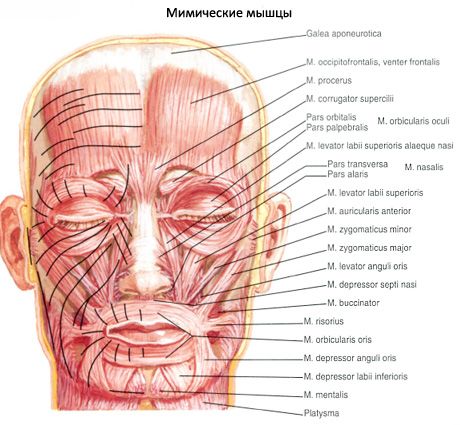
The muscle that lowers the septum of the nose (m.depressor septi nasi) is often part of the alar part of the nasal muscle. The bundles of this muscle begin above the medial incisor of the upper jaw and are attached to the cartilaginous part of the nasal septum.
Function: pulls the nasal septum downwards.
Innervation: facial nerve (VII).
Blood supply: superior labial artery.
 [
[ Where does it hurt?