Medical expert of the article

New publications
The muscles surrounding the eye socket
Last reviewed: 07.07.2025

All iLive content is medically reviewed or fact checked to ensure as much factual accuracy as possible.
We have strict sourcing guidelines and only link to reputable media sites, academic research institutions and, whenever possible, medically peer reviewed studies. Note that the numbers in parentheses ([1], [2], etc.) are clickable links to these studies.
If you feel that any of our content is inaccurate, out-of-date, or otherwise questionable, please select it and press Ctrl + Enter.
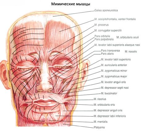
The eye slit is surrounded by bundles of the orbicularis oculi muscle, which has several parts.
The circular muscle of the eye (m.orbicularis oculi) is flat, occupies the periphery of the orbit, is located in the thickness of the eyelids, and partially extends into the temporal region. The lower bundles of the muscle continue into the cheek area. The muscle consists of 3 parts: the eyelid, orbital, and lacrimal.
The palpebral part (pars palpebralis) is represented by a thin layer of muscle bundles that originate on the medial palpebral ligament and adjacent areas of the medial wall of the orbit. The muscle bundles of the palpebral part pass along the anterior surface of the cartilages of the upper and lower eyelids to the lateral corner of the eye; here the fibers intertwine, forming the lateral suture of the eyelid. Some of the fibers are attached to the periosteum of the lateral wall of the orbit.
The orbital part (pars orbitalis) is significantly thicker and wider than the palpebral part. It begins on the nasal part of the frontal bone, on the frontal process of the maxilla and the medial ligament of the eyelid. The bundles of this muscle pass outward to the lateral wall of the orbit, where the upper and lower parts continue into each other. The bundles of the frontal belly of the occipitofrontalis muscle and the muscle that wrinkles the eyebrow are woven into the upper part.
The lacrimal part (pars lacrimalis) originates on the lacrimal crest and the adjacent part of the lateral surface of the lacrimal bone. The fibers of the lacrimal part pass laterally behind the lacrimal sac and are woven into the wall of this sac and into the palpebral part of the orbicularis oculi muscle.
Function: The orbicularis oculi muscle is a sphincter of the palpebral fissure. The palpebral part closes the eyelids. When the orbital part contracts, folds are formed on the skin in the eye socket area. The greatest number of fan-shaped diverging folds are observed on the side of the outer corner of the eye. The same part of the muscle moves the eyebrow down, simultaneously pulling the skin of the cheek up. The lacrimal part expands the lacrimal sac, thereby regulating the outflow of tear fluid through the nasolacrimal duct.
Innervation: facial nerve (VII).
Blood supply: facial, superficial temporal, supraorbital and infraorbital arteries.
 [ 1 ]
[ 1 ]
Where does it hurt?