Medical expert of the article
New publications
Molluscum contagiosum
Last reviewed: 04.07.2025

All iLive content is medically reviewed or fact checked to ensure as much factual accuracy as possible.
We have strict sourcing guidelines and only link to reputable media sites, academic research institutions and, whenever possible, medically peer reviewed studies. Note that the numbers in parentheses ([1], [2], etc.) are clickable links to these studies.
If you feel that any of our content is inaccurate, out-of-date, or otherwise questionable, please select it and press Ctrl + Enter.
Causes molluscum contagiosum
The causative agent of the disease is the molluscus contagiosum virus, which is considered pathogenic only for humans and is transmitted either through direct contact (in adults - often during sexual intercourse) or indirectly through the use of common hygiene items (washcloths, sponges, towels, etc.).
The incubation period varies from several weeks to several months. Sometimes the disease occurs in people with reduced immunity, severe systemic diseases.
The virus (MCV) is an unclassified type of smallpox virus. The disease is widespread and affects only humans. Children under one year of age rarely get sick, possibly due to immunity acquired from the mother and a long incubation period.
According to numerous observations, molluscum contagiosum is more common in patients suffering from atopic dermatitis and eczema. This is due to both decreased skin reactivity and long-term use of topical steroids. Unusually widespread rashes have been noted in patients with sarcoidosis, in patients receiving immunosuppressive therapy, and in HIV-infected patients. Thus, cell-mediated immunity is of great importance in the occurrence and development of the infectious process.
Pathogenesis
The pathogenesis links have not been sufficiently studied, but the decisive role is played by the disruption of the epidermal growth factor. The virus penetrates into the keratinocytes of the basal layer of the epidermis and significantly increases the rate of cell division. Then, in the spinous layer, there is an active accumulation of viral DNA. As a result, a nodule is formed, in the center of which destruction and disintegration of epidermal cells occurs, while the cells of the basal layer are not affected. Thus, the central part of the nodule is represented by detritus containing hyaline bodies (mollusk bodies) with a diameter of about 25 μm, which, in turn, contain masses of viral material. Inflammatory changes in the dermis are insignificant or absent, but in the case of long-standing elements they can be represented by a chronic granulomatous infiltrate.
How is molluscum contagiosum transmitted?
Molluscum contagiosum is spread through direct contact with broken skin or contaminated objects. The main ways molluscum contagiosum is spread are:
- The main way molluscum contagiosum is spread is through direct physical contact with an infected person. This can include skin-to-skin contact with the rash, hand-to-hand contact, kissing, or sexual contact.
- Shared objects: The molluscum contagiosum virus can also be spread through shared objects such as towels, clothing, toys, pools, or showers that have been used by infected people. The virus can remain on these objects and be transmitted to other people when they are used.
- Scratching and trauma: The affected areas of skin can become a source of the virus through mechanical action, such as scratching, scraping or removing the rash. This can lead to the spread of the virus to other areas of the skin.
- Autoinfection: In rare cases, molluscum contagiosum can be spread from one part of the body to another in the same person. This can happen if infected skin comes into contact with healthy skin.
It is important to emphasize that molluscum contagiosum can be easily transmitted, especially among children and in situations where people are close to each other or use common hygiene items. Therefore, it is important to follow hygiene precautions and avoid direct contact with affected skin to prevent transmission of the virus.
Symptoms molluscum contagiosum
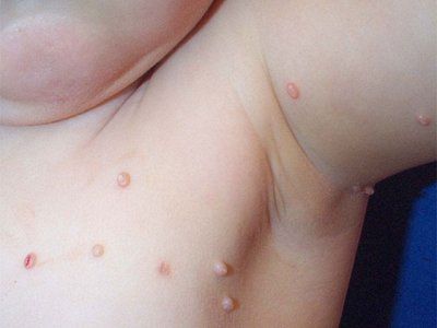
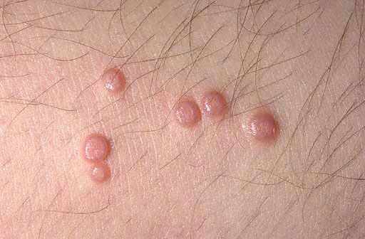
Molluscum contagiosum has an incubation period of 14 days to 6 months. The rash is represented by shiny pearly-white hemispherical papules with an umbilicated depression in the center. Slowly increasing in size, the papule can reach 5-10 mm in diameter in 6-12 weeks. In solitary lesions, the papule diameter reaches significant sizes. After injury or spontaneously, after several months, the papules can suppurate and ulcerate. Usually, after 6-9 months, the rash resolves spontaneously, but some persist for up to 3-4 years. The rash is most often localized on the face, neck, trunk, especially in the armpits, with the exception of sexually transmitted infections, when the anogenital area is usually affected. Elements can also be localized on the scalp, lips, tongue, mucous membrane of the cheeks, on any part of the skin, including an atypical localization - the skin of the soles. Papules can be localized on scars, tattoos.
At the sites of virus introduction, single or multiple, dense, shiny, painless, pink or grayish-yellow nodules appear, the size of which varies from a millet grain to a pea. There is a characteristic depression in the center of the element. In children, they are most often located on the face, neck, back of the hands, and can be randomly scattered over the entire skin or grouped into separate foci.
In children under 10 years of age, molluscum contagiosum is most often localized on the face. Here, rashes are most often located on the eyelids, in particular, along the eyelash line, around the eyes, on the nose and around it, on the cheeks, chin. In addition to the face, other areas are often affected - the submandibular, neck, chest, upper limbs, trunk, external genitalia, etc.

If in children frequent localization on the face (about 1/2 of all cases) is explainable and is a common occurrence, then molluscum contagiosum in adults is rarely located on the face and is considered a result of weakened immunity (atopy, immunosuppressive therapy, AIDS, etc.). Adults are considered immune to the virus, so its rapid spread over the skin, especially on the face, as well as the appearance of atypical forms indicate acquired immunodeficiency. In such cases, it is necessary to clarify the anamnesis, conduct the necessary studies (including HIV infection) to clarify the pathogenesis.
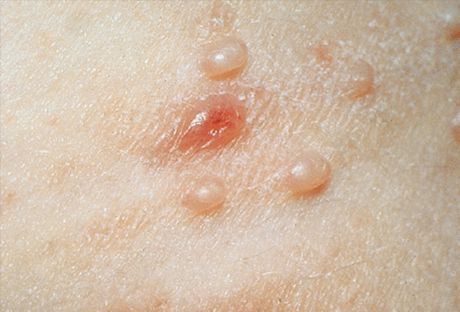
In typical cases, the primary elements of the rash are non-inflammatory, semi-translucent nodules, whitish-matte, flesh-colored or yellowish-pink, the size of a pinhead or millet grain. More often, there are several such elements, they are located in small groups, asymmetrically and do not cause subjective sensations. Small elements do not show umbilical depressions in the center and they are very reminiscent of milia or young forms of flat warts. The number and size slowly increase, and they reach an average size of a pea. Such elements have a hemispherical shape, a characteristic depression in the center, and a dense consistency. When squeezing the nodule from the sides with tweezers, a whitish mushy mass is released from the umbilical depression, consisting of keratinized epidermal cells, molluscum bodies and fat. This helps in clinical and microscopic diagnosis.
Clinical manifestations on the face can be quite diverse and resemble some other dermatoses with similar manifestations. In addition to the typical elements described above, atypical forms can also be encountered. In cases where an individual element reaches a size of 1 cm or more, a giant form similar to a cyst is noted. Some elements (usually giant) ulcerate and resemble keratoacanthoma, ulcerated basalioma or squamous cell skin cancer. Individual elements can become inflamed, suppurate, therefore they change their appearance and become similar to acne (acneiform), elements of chickenpox (varicelliform), folliculitis (folliculitis-like) or furuncle (furuncle-like). Such clinical forms present certain difficulties for diagnosis. The simultaneous presence of typical nodules facilitates diagnosis. Suppuration usually ends with spontaneous regression of this element.
In HIV-infected individuals, the rashes are multiple, localized mainly on the face and are resistant to traditional therapy.
In adults, with sexually transmitted infections, rashes can be localized on the genitals and in the perigenital areas.

A characteristic feature of the nodules is the release of a white mushy mass from the central depression of the papules when they are squeezed with tweezers. Subjective sensations are usually absent. Sometimes the rashes can merge into large uneven tumor-like formations ("giant molluscum") or disappear spontaneously.
Histopathology
A characteristic formation consisting of pear-shaped lobules is observed. The epidermal cells are enlarged, there are many intraplasmic inclusions (mollusk bodies) that contain viral particles. There is a small inflammatory infiltrate in the dermis.
The diagnosis is based on the characteristic clinical picture. The diagnosis can be verified by detecting characteristic "mollusk bodies" during a microscopic examination of the mushy mass squeezed out of the mollusk (shiny when viewing the native preparation in a dark field of the microscope or stained dark blue with the main saviors - methylene blue or Romanovsky-Giemsa). In some cases, a histological examination of the affected skin is carried out to clarify the diagnosis.
Diagnostics molluscum contagiosum
On the face of children and adolescents, molluscum contagiosum is primarily differentiated from flat warts, milia, angiofibromas (isolated and symmetrical), syringoma, epidermodysplasia verruciformis, Darier's disease, trichoepithelioma, and atypical forms - from cysts, acne, chickenpox rashes, folliculitis, furuncle, barley.
In middle-aged and elderly people, in addition to the above-mentioned dermatoses, which are rarer for this group, the differential diagnosis of molluscum contagiosum is carried out with senile hyperplasia of the sebaceous glands, xanthelasma, papular xanthoma, nodular elastoidosis with cysts and comedones (Favre-Racouchot disease), with hydrocystoma (on the eyelids), keratoacanthoma, ulcerated basalioma or squamous cell carcinoma of the skin.
What do need to examine?
Who to contact?
Treatment molluscum contagiosum
Patients should avoid visiting swimming pools, public baths, and carefully observe personal hygiene rules. Any cosmetic procedures are undesirable. There is no specific treatment for molluscum contagiosum.
Medicines
Treatment for molluscum contagiosum may involve the use of various medications. Here are some of them:
- Topical retinoids: Examples of these medications include Tretinoin (Retin-A) and Tazarotene (Tazorac). These medications can help speed up the drying process of molluscum.
- Trichloroacetic acid (TCA): This chemical can be applied directly to mollusks to remove them. The procedure must be performed by a doctor.
- Imiquimod (Aldara): This cream can be used to stimulate the immune system to kill molluscum contagiosum virus cells.
- Subcutaneous imiquimod (Zyclara): This drug is similar to Aldara cream, but is given as an injection under the skin.
- Cantharidin: This chemical can be used to treat molluscum contagiosum, but it must be applied by a doctor because it can cause skin irritation.
How to remove molluscum contagiosum?
You can remove molluscum contagiosum with epilation tweezers and scraping with a spoon, followed by lubricating the erosion with a 1% alcohol solution of iodine. Before removal, local anesthesia with a 10% lidocaine spray or short-term freezing with liquid nitrogen is recommended (especially in children). Such treatment does not leave permanent marks. Diathermocoagulation, cryo- or laser destruction on the face is best avoided, as they can leave cicatricial changes. In small children, in some cases, it is advisable to leave the elements without treatment or limit yourself to long-term external use of interferon ointment.
Patients (or parents of children) should be aware of possible relapses of the disease, so all family members, as well as the patient, should be examined 2-3 weeks after completion of treatment, and the identified predisposing factors should be taken into account.
It is necessary to remove the nodes from the Volkman spoon, diathermocoagulation followed by lubrication with a 2-5% alcohol solution of iodine. Diathermocoagulation of elements is also possible. In disseminated forms of the disease, antiviral agents are used: proteflazit (15-20 drops 2 times a day for adults), interferon (3-4 drops in the nose 4-5 times a day) or methisazone orally.
Clinical guidelines
Molluscum contagiosum is a viral disease caused by the Molluscum contagiosum virus. It appears as small, round, smooth, papular lesions on the skin that may be white, pink, or firm. The virus is spread by contact with infected patients or objects such as towels or clothing.
Clinical guidelines for the treatment of molluscum contagiosum may include the following:
- Observation without treatment: In some patients, molluscum may resolve on its own within a few months to a few years. This observational approach may be suggested for children and adults with a small number of lesions.
- Extrusion (extraction): This is a procedure in which a doctor uses an instrument to squeeze out the contents of the molluscum. Extrusion is usually performed by a doctor or dermatologist and can be painful. It can be effective, but burns, scarring, or recurrence may occur.
- Chemical treatments: Your doctor may prescribe chemicals, such as trichloroacetic acid (TCA) or subcutaneous imiquimod, to help remove molluscum. These treatments may also cause redness, burning, and scaling.
- Surgical treatment: Surgical removal of molluscum using scissors, laser, or electrocautery may be considered in cases where other methods are ineffective.
- Preventing Spread: Because molluscum contagiosum is easily spread, it is important to avoid contact with infected skin areas and objects that may be contaminated with the virus.
Treatment of molluscum contagiosum should be carried out under the supervision of a doctor, and the choice of method depends on the number and location of molluscum, as well as the age and general condition of the patient. For specific recommendations and prescriptions, you should always consult a medical specialist.
List of some books and studies related to the study of molluscum contagiosum
- "Molluscum Contagiosum: Diagnosis and Clinical Management" Author: John Bordeau, MD Year of publication: 2012
- "Molluscum Contagiosum: A Medical Dictionary, Bibliography, and Annotated Research Guide to Internet References" Author: Health Publica Icon Health Publications Year of publication: 2004
- "Molluscum Contagiosum: The Complete Guide" Author: Frederick Babinski, MD Year of publication: 2017
- "Epidemiology of Molluscum Contagiosum in Children: A Systematic Review" Authors: Seyed Alireza Abtahi-Naeini, Mahin Aflatoonian, and others Year of publication: 2015
- "Molluscum Contagiosum Virus: Current Trends and Future Prospects" Authors: Anubhav Das, AK Singh, and others Year of publication: 2019
- "Molluscum Contagiosum Virus: The Neglected Cousin of Poxviruses" Authors: SR Patel, G. Varveri, and others Year of publication: 2019
Literature
Butov, Yu. S. Dermatovenereology. National leadership. Brief edition / ed. Yu. S. Butova, Yu. K. Skripkina, O. L. Ivanova. - Moscow: GEOTAR-Media, 2020.


 [
[