Medical expert of the article

New publications
Skiascopy
Last reviewed: 07.07.2025

All iLive content is medically reviewed or fact checked to ensure as much factual accuracy as possible.
We have strict sourcing guidelines and only link to reputable media sites, academic research institutions and, whenever possible, medically peer reviewed studies. Note that the numbers in parentheses ([1], [2], etc.) are clickable links to these studies.
If you feel that any of our content is inaccurate, out-of-date, or otherwise questionable, please select it and press Ctrl + Enter.
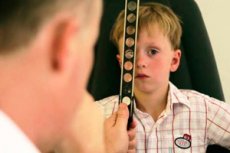
Skiascopy (from the Greek scia - shadow, scopeo - I examine) is a method of objectively studying clinical refraction, based on observing the movement of shadows obtained in the area of the pupil when the latter is illuminated using various techniques.
Without delving into the essence of the physical phenomena on which skiascopy is based, the main position of this method can be formulated as follows: the movement of the shadow is not observed if the further point of clear vision coincides with the source of illumination of the pupil, i.e., in fact, with the position of the researcher.
Methodology of implementation
Skiascopy is performed using the following technique.
The doctor sits opposite the patient (usually at a distance of 0.67 or 1 m), illuminates the pupil of the eye being examined with the ophthalmoscope mirror and, turning the device around the horizontal or vertical axis in one direction or the other, observes the nature of the shadow movement against the background of the pink reflex from the fundus in the pupil area. During skiascopy with a flat mirror from a distance of 1 m in the case of hyperopia, emmetropia and myopia less than -1.0 D the shadow moves in the same direction as the mirror, and with myopia greater than -1.0 D - in the opposite direction. When using a concave mirror, the ratios are reversed. The absence of movement of the light spot in the pupil area during skiascopy from a distance of 1 m using both a flat and a concave mirror indicates that the person being examined has myopia of -1.0 D.
This method is used to determine the type of refraction. To determine its degree, the shadow movement neutralization method is usually used. For myopia greater than -1.0 Dptr, negative lenses are applied to the eye being examined, first weak and then stronger (in absolute value) until the shadow movement in the pupil area stops. In cases of hypermetropia, emmetropia, and myopia less than -1.0 Dptr, a similar procedure is performed with positive lenses. For astigmatism, the same is done separately in the two main meridians.
The required refraction value can be determined using the following formula:
R= C-1/D.
Where R is the refraction of the eye being examined (in diopters: myopia - with a "-" sign, hyperopia - with a "+" sign; C is the power of the neutralizing lens (in diopters); D is the distance from which the examination is performed (in meters).
Some practical recommendations for performing skiascopy can be formulated as follows.
- It is recommended to use an electroskiascope, i.e. a device with a built-in light source, if possible, or, if it is not available, a flat ophthalmoscopic mirror and an incandescent lamp with a transparent bulb (smaller area of the light source). When examining with a flat mirror (compared to a concave one), the shadow is more pronounced and homogeneous, its movements are easier to assess, and smaller turns of the mirror are required to move the shadow.
- To neutralize the shadow, one can use either special skiascopic rulers or lenses from a set, which are inserted into a trial frame. The advantage of the latter method, despite the increase in examination time, is associated with the precise observance of a constant distance between the lenses and the apex of the cornea, as well as the possibility of using cylindrical lenses to neutralize the shadow in astigmatism (the cylindroskiascopy method). The use of the first method is justified when examining children, since in these cases the doctor, as a rule, is forced to hold the skiascopic rulers in front of the patient's eye.
- It is advisable to perform skiascopy from a distance of 67 cm, which is easier to maintain during the examination, especially when determining refraction in young children.
- When examining the eye under cycloplegia, the subject should look at the mirror opening, and in cases of intact accommodation, past the doctor's ear on the side of the eye being examined.
- When using a skiascopic ruler, you should try to keep it vertical and at a standard distance from the eye (approximately 12 mm from the top of the cornea).
If there is no movement of the shadow when changing a row of lenses, the arithmetic mean value of the power of these lenses should be taken as the indicator for calculations.
When performing skiascopy under conditions of drug-induced cycloplegia, which, as noted, is accompanied by pupil dilation (mydriasis), the following difficulties are possible. The shadow can move in different directions, and shadow neutralization is provided by different lenses in different areas of the pupil - the so-called scissors symptom. This fact indicates irregular astigmatism, most often caused by a non-spherical shape of the cornea (for example, in keratoconus - corneal dystrophy, accompanied by a change in its shape). In this case, the diagnosis is clarified using an ophthalmometer. If any pattern in the movement of the shadow is established, for example, a different character in the center and on the periphery of the pupil, then this movement should be neutralized, focusing on the movement of the shadow in the central zone.
An unstable, changing nature of the shadow movement during the examination usually indicates insufficient cycloplegia and the possible influence of accommodation tension on the results of skiascopy.
Difficulties may arise during a skiascopic examination of an eye with low visual acuity and, as a consequence, unstable non-central fixation. As a result of the constant movement of this eye during the examination, the refraction of not the macula but other non-central areas of the retina will be determined. In such cases, an object is presented to the leading eye for fixation, it is moved and, using combined movements, the poorly seeing eye is set in a position in which the light block of the ophthalmoscope or skiascope is located in the center of the cornea.
To clarify refraction in astigmatism, you can use line-skiascopy, or strip skiascopy. The study is carried out using special skiascopes that have a light source in the form of a strip that can be oriented in different directions. Having installed the light strip of the device in the desired position (so that it does not change when moving to the pupil), skiascopy is carried out according to the general rules in each of the main meridians found, achieving a cessation of the movement of the strip shadow.
Cylindroskiascopy
Cylindroskiascopy allows to specify the data obtained during skiascopy. First, a regular skiascopy with rulers is performed, the position of the main meridians of the astigmatic eye and the power of the lenses, when used, the movement of the shadow in each of them is approximately determined. A trial frame is put on the patient and a spherical and astigmatic lens, which should ensure the cessation of shadow movement simultaneously in both main meridians, are placed in the socket located opposite the eye being examined, and skiascopy is performed in them. The cessation of shadow movement in one or the other direction indicates that the skiascopic refraction indices have been determined correctly. If the shadow moves not in the direction of the cylinder axis or its active section, but between them (usually at an angle of approximately 45° to them), then the axis of the cylinder is installed incorrectly. In this case, the cylinder placed in the frame is rotated until the direction of shadow movement coincides with the direction of the axis.
The main advantage of skiascopy is its availability, since no complex equipment is required to perform the examination. However, certain skills, experience and qualifications are required to perform skiascopy. In addition, in some cases (for example, with astigmatism with oblique axes), the information content of the technique may be limited.