Medical expert of the article
New publications
Meningioma of the optic nerve sheath
Last reviewed: 07.07.2025

All iLive content is medically reviewed or fact checked to ensure as much factual accuracy as possible.
We have strict sourcing guidelines and only link to reputable media sites, academic research institutions and, whenever possible, medically peer reviewed studies. Note that the numbers in parentheses ([1], [2], etc.) are clickable links to these studies.
If you feel that any of our content is inaccurate, out-of-date, or otherwise questionable, please select it and press Ctrl + Enter.

Symptoms of Optic Nerve Sheath Meningioma
The disease manifests itself in middle age with a gradual unilateral decrease in vision. Temporary visual impairment may be the first symptom.
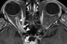
The classic triad is: decreased vision, optic nerve atrophy, and opticociliary vascular shunts. However, the simultaneous appearance of all three signs is rare. The sequence of signs is as follows:
- Dysfunction of the optic nerve and chronic stagnation of the disc, followed by atrophy.
- Opticociliary vascular shunts, which are found in approximately 30% of cases, regress with the development of optic nerve atrophy.
- Limited mobility, especially upward, since the tumor can “split”) the optic nerve.
- Exophthalmos appears due to tumor growth within the muscular funnel and develops after a decrease in vision.
The sequence is the opposite of that seen in tumors growing outside the dura mater, where exophthalmos appears long before compression of the optic nerve.
What's bothering you?
What do need to examine?
Who to contact?
Treatment of optic nerve sheath meningioma
- Monitoring of middle-aged patients with slow tumor growth, as the prognosis is good.
- Surgical removal in young patients with aggressive tumors, especially in a blind eye.
- Radiation in some cases.


 [
[