Medical expert of the article

New publications
Infectious erythema
Last reviewed: 12.07.2025

All iLive content is medically reviewed or fact checked to ensure as much factual accuracy as possible.
We have strict sourcing guidelines and only link to reputable media sites, academic research institutions and, whenever possible, medically peer reviewed studies. Note that the numbers in parentheses ([1], [2], etc.) are clickable links to these studies.
If you feel that any of our content is inaccurate, out-of-date, or otherwise questionable, please select it and press Ctrl + Enter.
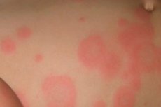
When affected by various infections, focal redness may appear on the skin - infectious erythema, which is a sign that the infection has caused a reaction in the form of increased blood flow to the skin area.
Due to the lack of terminological clarity in dermatology, erythema can be used to refer to some conditions with red spots on the skin. [ 1 ]
Epidemiology
Medical statistics cannot record cases of skin redness as a symptom of infectious skin lesions or systemic diseases, but it tracks data on the etiological factors of alteration.
Thus, one of the most common causes of infectious erythema is streptococcal infection, which accounts for almost half of cases of infectious erythema nodosum in children and more than 40% of cases in adults. [ 2 ]
In 20% of cases of parvovirus B19 infection in children and adults, there are no symptoms. And in cases of tick bites, characteristic erythema is observed in eight out of ten cases. [ 3 ], [ 4 ]
Parvovirus B19 infection in pregnant women can cause severe complications in the fetus. These complications include miscarriage, intrauterine death, and hydrops fetalis.[ 5 ] The risk of fetal loss after acute infection is approximately 5%. Mothers in the second trimester of pregnancy are at greatest risk for complications from parvovirus B19, but cases have been reported at all stages of pregnancy.[ 6 ]
Patients with sickle cell or other chronic hemolytic diseases may be more severely affected than other populations. [ 7 ] Parvovirus B19 infection destroys reticulocytes. This causes a decrease or temporary cessation of erythropoiesis. These individuals may develop aplastic crisis and develop severe anemia. Often these patients will be much sicker with fever, malaise, and lethargy. Patients with aplastic crisis will have pallor, tachycardia, and tachypnea due to severe anemia. [ 8 ]
Causes infectious erythema
Any redness of the skin (erythros means red in Greek) is a natural cause for concern, but it is a special case when the causes of erythema are related to infections.
An example is skin damage caused by Streptococcus pyogenes bacteria, a group A beta-hemolytic streptococcus, which leads to the development of various types of streptoderma, as well as erysipelas.
The reaction in the form of reddening of the skin can be caused by the bacteria Staphylococcus aureus, Mycoplasma hominis, Yersinia enterocolitica, Erysipelothrix rhusiopathiae, as well as the herpes virus (including type IV - Epstein-Barr virus), erythroparvovirus (Primate erythroparvovirus 1). It is assumed that among the causes of persistent elevated erythema, which appears in the joint area with inflammation of the walls of skin capillaries (vasculitis), there may be an immune reaction to the bacteria Streptococcus spp. and Escherichia coli (E. coli).
Infectious-allergic erythema refers to allergic dermatoses. It can also be complicated by infection microbial eczema or skin vasculitis of allergic and infectious origin.
In cancer patients, systemic erythema may be due to bacterial infection, most commonly Streptococcus viridans and Arcanobacterium haemolyticum.
Infectious erythema in adults and children occurs with skin lesions caused by arthropods, primarily by the ixodid tick, which carries the bacteria Borrelia burgdorferi [ 9 ] - the cause of Lyme disease, which begins with the appearance of redness at the site of the bite - erythema migrans chronica Afzelius-Lipschütz. [ 10 ], [ 11 ]
Risk factors
The main risk factors, according to experts, include a decrease in general and local immunity (and, accordingly, all conditions and pathologies that cause immunosuppression), foci of chronic bacterial or viral infection in the body - streptococcal, staphylococcal, herpesvirus, as well as increased sensitivity (sensitization) - with a tendency to allergic reactions.
Pathogenesis
The pathogenesis of the development of infectious erythema, as one of the types of red spots on the skin of the body, is caused by increased blood flow in the superficial capillaries during an inflammatory reaction, which, in essence, is protective and aimed at neutralizing antigens and toxins produced by pathogenic microorganisms. [ 12 ]
What mediators trigger the defense mechanism and what immune cells ensure its functioning are described in detail in the publication – Systemic inflammatory response syndrome.
For more information on how the most common bacterial and viral infections work, see the articles:
Symptoms infectious erythema
It is necessary to pay attention to the types of infectious erythema, which are nosological units, and are also traditionally distinguished by most dermatologists as separate diseases, but are not registered in the International Classification of Diseases.
Erythema multiforme infectiosum
Erythema exudative multiforme, erythema multiforme Hebra (named after the 19th century Austrian dermatologist F. von Hebra who first described it), multiform or infectious erythema multiforme (code L51 according to ICD-10) is considered a cutaneous immune reaction to an infection (as well as a number of drugs). Often, this erythema is part of a specific response to the activation of the herpes simplex virus (HSV types I and II): in half of the cases, patients have a history of periodic herpetic eruptions on the lips.
As a rule, the incubation period of erythema multiforme does not exceed 48 hours, and its first signs are the appearance of convex rounded reddenings with clearly defined contours on the skin of the extremities, small at first, but quickly increasing (up to 30 mm in diameter). Then the erythema spreads to the upper part of the body and face, and in the center of the spots the hyperemia may become more intense; the presence of pustules (blisters filled with serous fluid) or crusts is possible. Itching may occur. Usually, the rash goes away after two to four weeks. [ 13 ]
In cases of severe erythema multiforme, Stevens-Johnson syndrome develops with high fever, headache and joint pain, ulcers on the oral mucosa and genitals, redness of the eyes and their increased sensitivity to light.
Read also – Erythema multiforme exudative. Causes, symptoms, diagnosis, treatment
Infectious erythema nodosum
In addition to the fact that this type of skin redness is one of the symptoms of the secondary focal form of such a zoonotic disease as pseudotuberculosis, the causative agent of which is the enterobacterium Yersinia pseudotuberculosis, infectious erythema nodosum has the ICD-10 code L52. [ 14 ]
It is associated with a bacterial infection - streptococcal or tuberculosis, as well as a viral infection (infectious mononucleosis), and typical symptoms include fever, painful firm nodules in the skin and surrounding swollen red spots on the skin of the front of the shins, joint pain.
The nodules may become inflamed and then flatten and disappear, leaving behind hematomas or depressions in the skin – like a mark left by damage to the subcutaneous tissue. [ 15 ]
Redness may disappear on its own within three to six weeks.
Infectious erythema of Rosenberg
Rosenberg's macular (spotted) erythema (described by Russian infectious disease specialist N. Rosenberg) suddenly appears in adolescence and young adulthood. The pathological condition manifests itself with fever and chills, as well as headache and aching joints. Rashes on the legs, arms and trunk appear approximately four to five days later - as separate red round spots.
The spots are characterized by a rapid increase in diameter (sometimes three to five times) and their merging to form large hyperemic zones, which dermatologists call erythematous fields. The color of the rash fades after three days, and after a few more days they disappear, and in their place peeling of the epidermis may be observed. As experience shows, the duration of sudden spotted erythema varies from one to two weeks.
Infectious toxic erythema
According to ICD-10, toxic erythema has the code L53. In clinical practice, such systemic erythema is observed in toxic shock syndrome caused by group A streptococcal infection. For more information, see – Symptoms of streptococcal infection.
In addition, staphylococcal infection, primarily Staphylococcus aureus toxins, may be involved in the development of toxic shock – with diffuse erythema of the trunk and arms, fever, drop in blood pressure, muscle pain and loss of consciousness.
Almost half of infants develop toxic erythema of the newborn (Erythema toxicum neonatorum, ICD-10 code P83.1) on the second to fifth day after birth. These are red spots on the skin with white or yellow nodules (or fluid-filled blisters) that disappear within one to two weeks. Officially, this condition is considered liopathic, but many researchers explain its etiology by the activation of the immune system of newborns in response to the formation of skin microflora in the neonatal period.
For more details see – Erythema of the skin of newborns: causes, consequences, treatment
Erythema infectiosum in children – fifth disease
What is fifth disease or Chamer's erythema infectiosum? It is a viral infection characterized by skin lesions (ICD-10 code B08.3); the causative agent is parvovirus infection - erythrovirus (parvovirus) B19, which is now simply called the B19 virus of the genus Erythroparvovirus, which is transmitted by airborne droplets. [ 16 ]
It is common in children aged 5-15 (especially in winter and spring), but adults can also get sick. The incubation period is from four days to two weeks, and the child is contagious before a rash appears on the skin.
At the end of the 19th century, the disease was described by the Austrian doctor A. Tschamer, who considered it a variant of German measles (rubella), and the redness on the cheeks was called Tschamer's erythema. And the fifth disease, because it was the fifth in the list of six most common pediatric infectious diseases accompanied by a rash.
For more information, see – Erythema infectiosum: Parvovirus B19 antibodies in the blood
Initial symptoms resemble flu and include headache, body aches, fever, and chills; the throat may ache. In adults, there is no rash or erythema (but joints may hurt), and in children, after two to three days, a bright red rash appears on the cheeks, sometimes red mesh rashes on the limbs and torso, which can last from 10 days to several weeks.
Complications and consequences
Complications of severe cases of erythema multiforme infectious include scarring, focal inflammation of the subcutaneous tissue, eye damage, and inflammation of internal organs. [ 17 ]
Also read – Consequences and complications of streptoderma
Erythema in Lyme disease may be complicated by the development of local skin atrophy.
Infection with parvovirus 19 in people with weakened immune systems or hematological diseases can lead to bone marrow damage and cause severe anemia. And in pregnant women infected before the 20th week, there is a risk of fetal death. [ 18 ]
Diagnostics infectious erythema
Clinical diagnosis of infectious diseases and conditions in which erythema appears on the skin involves a complete patient history, including medications taken, recent travel, bites and other factors, as well as examination of the skin, including the characteristics of the rash itself (localization, morphological features, etc.). The same diagnostic technique is used for erythemas, which are identified as independent nosological units (although in almost half of the cases, doctors cannot determine their causes).
Blood tests, in addition to general and biochemical, include determination of bacterial antigens (IgA, IgG, IgM) in the serum of the crocodile, analysis for Staphylococcus aureus and antistreptococcal antibodies, analysis for herpes, etc. A biopsy of the affected skin is necessary for erythema nodosum. [ 19 ]
Instrumental diagnostics is carried out using dermatoscopy.
Differential diagnosis
The main problem is differential diagnosis of infectious erythema: with common dermatological diseases (dermatitis, pink lichen, erythrokeratoderma, fungal skin lesions), with allergic conditions (including drug toxicoderma), as well as with skin manifestations of childhood infections, systemic lupus erythematosus and other exanthemas (rashes) of various etiologies. For example, with erythematous skin lesions in Wagner's disease (dermatomyositis) or glucagonoma (pancreatic tumor). [ 20 ]
Who to contact?
Treatment infectious erythema
In some cases, such as parvovirus erythema in children and other rashes of viral origin, specific treatment is not required: it is enough to use non-steroidal anti-inflammatory drugs (NSAIDs) to relieve fever and headache. The exception is the herpes virus, for more details see - Treatment of simple herpes
What medications are most often used for bacterially caused skin redness? These are systemic antibiotics for streptoderma in children and adults; various topical agents:
- Ointments for streptoderma
- Ointments for eczema
- Ointments for skin rashes
- If the reddened skin itches, antihistamines can help relieve the itching.
Also read:
The development of toxic shock in systemic toxic erythema associated with streptococcal or staphylococcal infection is life-threatening and requires emergency medical care.
And first aid is necessary for bites from arthropods that are hematophagous; comprehensive information on what to do is in the material - Tick bites in humans.
Prevention
Non-specific preventive measures include good hygiene – frequent hand washing to reduce the risk of infection. You should also avoid contact with infected people.
Forecast
In the absence of complications, erythema infectiosum has a favorable prognosis. [ 21 ] Symptoms of erythema infectiosum are usually self-limited in immunocompetent patients. These symptoms are usually mild, and some people may be asymptomatic. In immunocompromised patients or in patients with hematologic disorders, symptoms may be more severe. Chronic infection and chronic anemia may occur in immunocompromised individuals. Acute infection and exposure to the fetus can be fatal. The risk of fetal death is highest in infected pregnant women under 20 weeks of age.