Medical expert of the article

New publications
Erythema multiforme exudative: causes, symptoms, diagnosis, treatment
Last reviewed: 04.07.2025

All iLive content is medically reviewed or fact checked to ensure as much factual accuracy as possible.
We have strict sourcing guidelines and only link to reputable media sites, academic research institutions and, whenever possible, medically peer reviewed studies. Note that the numbers in parentheses ([1], [2], etc.) are clickable links to these studies.
If you feel that any of our content is inaccurate, out-of-date, or otherwise questionable, please select it and press Ctrl + Enter.
Erythema multiforme exudative is an acute, often recurrent disease of the skin and mucous membranes of infectious-allergic genesis, a polyetiological disease, mainly of toxic-allergic genesis, most often developing under the influence of infections, especially viral ones, and the effects of drugs. This disease was first described by Hebra in 1880.
Causes exudative erythema multiforme
The causes and pathogenesis of erythema multiforme exudative remain unclear. However, many scientists believe that the disease has a toxic-allergic genesis. The disease is considered a hypereric reaction directed at keratinocytes. Circulating immune complexes are detected in the blood serum of patients, and deposition of IgM and C3 components of the complement in the blood vessels of the dermis is noted. Trigger factors can be viral and bacterial infections, drugs. A connection with rickettsiosis has been noted. There are two forms of the disease: idiopathic with unknown etiology and secondary with an identified etiological factor.
Pathogenesis
Histopathology of erythema multiforme exudative. The histopathological picture depends on the clinical nature of the rash. In the maculopapular form, spongiosis and intracellular edema are observed in the epidermis. Edema of the papillary layer and perivascular infiltrate are observed in the dermis. The infiltrate consists of lymphocytes and some polymorphonuclear leukocytes, sometimes eosinophils. In bullous rashes, blisters are localized under the epidermis and only in old rashes can they sometimes be detected intraepidermally. Acantholysis is always absent. Sometimes, erythrocyte extravasates are visible without signs of vasculitis.
Pathomorphology of erythema multiforme exudative. Characteristic changes in the epidermis and dermis, but in some cases the epidermis changes predominantly, in others - the dermis. In this regard, three types of lesions are distinguished: dermal, mixed dermo-epidermal and epidermal.
In the dermal type, infiltration of the dermis of varying intensity is observed, sometimes occupying almost its entire thickness. Infiltrates consist of lymphocytes, neutrophils and eosinophils, granulocytes. With pronounced edema of the papillary layer of the dermis, blisters may form, the cover of which is the epidermis together with the basal membrane.
The dermo-epidermal type is characterized by the presence of a mononuclear infiltrate located not only perivascularly, but also near the dermo-epidermal junction. Hydropic dystrophy is observed in the basal cells, and necrobiotic changes are observed in the spinous cells. In some areas, the infiltrate cells penetrate the epidermis and, as a result of spongiosis, can form intraepidermal vesicles. Hydropic dystrophy of the basal cells in combination with pronounced edema of the papillary layer of the dermis can lead to the formation of subepidermal vesicles. Quite often, this type produces extravasates of erythrocytes.
In the epidermal type, only weak infiltration is observed in the dermis, mainly around the superficial vessels. Even in the early stages, the epidermis contains groups of epithelial cells with necrotic phenomena, which then undergo lysis, merge into a solid homogeneous mass, separating with the formation of a subepidermal blister. This picture is similar to that in toxic epidermal necrolysis (Lyell's syndrome). Sometimes necrobiotic changes are observed in the superficial parts of the epidermis and, together with edema, lead to the formation of subkeratinous blisters with subsequent rejection of its upper parts. In these cases, it is difficult to differentiate exudative erythema multiforme from herpetiform dermatitis and bullous pemphigoid.
Histogenesis of erythema multiforme exudative. The underlying mechanism of disease development is most likely immune. Direct immunofluorescence microscopy reveals a high titer of intercellular circulating antibodies in patients, but the results of direct immunofluorescence microscopy of the affected tissue are negative. These antibodies are capable of fixing complement, unlike antibodies in pemphigus. Scientists have found an increase in the number of lymphokines, macrophage factor, which indicates a cellular immune response. In the cellular infiltrate in the dermis, predominantly T-helper lymphocytes (CD4+) are detected, and in the epidermis - predominantly cytotoxic T-lymphocytes (CD8+). Immune complexes are also involved in the pathogenesis, which is primarily manifested by damage to the walls of blood vessels in the skin. Thus, it is assumed that a combined immune reaction develops, including a delayed-type hypersensitivity reaction (type IV) and an immune complex allergic reaction (type III). An association of the disease with the HLA-DQB1 antigen has been revealed.
Symptoms exudative erythema multiforme
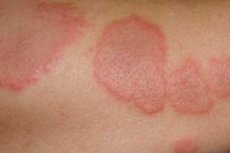
Clinically manifested by small erythematous edematous spots, maculopapular rashes with eccentric growth with the formation of two-contour elements due to the brighter peripheral and cyanotic central part. Ring-shaped, cockade-shaped figures, vesicles, and in some cases blisters with transparent or hemorrhagic contents, vegetations may occur. The preferred localization is the extensor surfaces, especially of the upper limbs. Rashes often occur on the mucous membranes, which is more typical of the bullous form of exudative erythema multiforme. The most severe clinical variety of the bullous form of the disease is Stevens-Johnson syndrome, which occurs with high fever, joint pain. There may be signs of myocardial dystrophy and damage to other internal organs (hepatitis, bronchitis, etc.). There is a pronounced tendency for erythema multiforme to relapse, especially in the spring and autumn.
In clinical practice, two forms of erythema multiforme exudative are distinguished - idiopathic (classical) and symptomatic. In the idiopathic form, it is usually impossible to establish the etiologic factor. In the symptomatic form, a certain factor causing the rash is known.
The idiopathic (classical) form usually begins with prodromal symptoms (malaise, headaches, fever). After 2-3 days, symmetrically located limited spots or flattened edematous papules of a round or oval shape, 3-15 mm in size, pink-red or bright red in color, appear acutely, increasing in size at the periphery. The peripheral ridge acquires a cyanotic tint, the central part sinks. In the center of individual rashes, new papular elements with exactly the same development cycle are formed. On the surface of the elements or on unchanged skin, various-sized vesicles, vesicles with serous or hemorrhagic contents, surrounded by a narrow inflammatory rim ("bird's eye symptom") appear. After some time, the blisters subside and their rim becomes cyanotic. In such areas, concentric figures are formed - herpes iris. Their dense cover opens up and erosions form, which quickly become covered with dirty, bloody crusts.
The preferred localization of the elements is the extensor surfaces of the upper limbs, mainly the forearms and hands, but they can also be located on other areas - the face, neck, shins, and the back of the feet.
Mucous membrane and lip lesions occur in approximately 30% of patients. At first, swelling and hyperemia occur, and after 1-2 days, blisters or bubbles appear. They quickly open, revealing bleeding bright red erosions, with the remains of the tires hanging along the edges. The lips swell, their red border is covered with bloody and dirty crusts and more or less deep cracks. Due to severe pain, eating can be very difficult. The outcome in most cases is favorable, the disease usually lasts 15-20 days and disappears without a trace, rarely, slight pigmentation remains in the places of rashes for some time. Sometimes the process can transform into Stevens-Johnson syndrome. The idiopathic form is characterized by seasonality of the disease (in the spring and autumn months) and relapses.
In the symptomatic form, rashes similar to classical exudative erythema appear. Unlike the classical type, the onset of the disease is associated with the intake of a certain agent, there is no seasonality, and the process is more widespread. In addition, the skin of the face and body is affected to no lesser extent, the cyanotic shade of the rash is not so pronounced, ring-shaped and "rainbow" (iris) figured rashes, etc. may be absent.
Drug-induced erythema multiforme exudative is mostly fixed in nature. Of the morphological elements, blisters predominate, especially when the process is localized in the oral cavity and on the genitals.
Depending on the clinical picture of the rash, there are macular, papular, maculopapular, vesicular, bullous or vesiculobullous forms of exudative erythema.
What do need to examine?
How to examine?
What tests are needed?
Differential diagnosis
Differential diagnosis includes fixed sulfanilamide erythema, disseminated lupus erythematosus, erythema nodosum, bullous pemphigoid, pemphigus, urticaria, and allergic vasculitis.
Treatment exudative erythema multiforme
In case of spotted, papular and mild bullous forms, symptomatic treatment is carried out - hyposensitizing (calcium preparations, sodium thiosulfate), antihistamines and externally - aniline dyes, corticosteroids. In severe cases, corticosteroids are prescribed orally (50-60 mg/day) or in the form of injections, in the presence of a secondary infection - antibiotics, herpes infection - antiviral drugs (acyclovir).
 [
[