Medical expert of the article

New publications
Hydropericardium
Last reviewed: 12.07.2025

All iLive content is medically reviewed or fact checked to ensure as much factual accuracy as possible.
We have strict sourcing guidelines and only link to reputable media sites, academic research institutions and, whenever possible, medically peer reviewed studies. Note that the numbers in parentheses ([1], [2], etc.) are clickable links to these studies.
If you feel that any of our content is inaccurate, out-of-date, or otherwise questionable, please select it and press Ctrl + Enter.
The pericardium is the fibrous membrane surrounding the heart – the pericardial sac, in the cavity of which, under the influence of various pathological factors, excess fluid can accumulate, which is diagnosed as hydropericardium, pericardial effusion (effusion) or dropsy of the pericardial sac. This condition can be life-threatening and requires detection and adequate treatment.
Non-inflammatory pericardial effusion has the ICD-10 code I31.3.
Epidemiology
According to foreign studies, among the causes of effusion in the pericardial cavity, 15-30% are pericarditis and various infections; 12-23% are oncology; 5-15% are connective tissue pathologies; 15-20% are iatrogenic causes.
In developing countries, more than 60% of cases of hydropericardium are caused by tuberculosis. In the presence of HIV, pericardial effusion is observed in, on average, a quarter of patients. Idiopathic hydropericardium accounts for up to half of cases.
In low birth weight neonates, the prevalence of pericardial fluid accumulation during parenteral nutrition via a central venous catheter is estimated at 1-3% (with mortality rates due to cardiac tamponade of up to 30-40%). [ 1 ]
Causes hydropericardium
Any accumulation of fluid in the body's cavities can be a sign of the disease. And the most common causes of hydropericardium include:
- inflammation of the pericardium - exudative, viral, and tuberculous pericarditis;
- congestive heart failure;
- acute myocardial infarction with development of Dressler syndrome; [ 2 ]
- viral myocarditis; [ 3 ]
- parasitic infection of the pericardium, for example, with trichinosis;
- autoimmune diseases such as rheumatic heart disease, rheumatoid arthritis, systemic lupus erythematosus (SLE);
- metastasis of lung cancer, breast cancer, melanoma, non-Hodgkin's lymphoma; [ 4 ]
- blunt and penetrating injuries to the heart area.
Hydropericardium is observed in pneumonia, especially if it is caused by mycoplasma or Haemophilus influenzae, with complications in the form of pleurisy, pericarditis or myocarditis.
Hydropericardium occurs with hypothyroidism - its myxedematous form and autoimmune thyroiditis.
Specialists observe a connection between hydropericardium and fluid accumulation in other cavities. In particular, effusion in one or both pleural cavities or hydrothorax and hydropericardium appear in cases of left-sided exudative pleurisy (especially tuberculous), pulmonary sarcoidosis, heart failure, myocarditis, SLE. chest injuries.
In patients with edematous syndromes - cardiac or nephrotic, as well as with liver cirrhosis, edema of the subcutaneous tissue - anasarca, hydropericardium and ascites - can simultaneously develop, that is, when fluid accumulates in the abdominal cavity in the form of peritoneal effusion.
Replacement of lung cells with connective tissue - pneumofibrosis and hydropericardium are most often associated with such an autoimmune disease as systemic scleroderma. Read more in the publication - Features of heart damage in systemic scleroderma
In addition, iatrogenic origin of fluid accumulation in the pericardium is possible: after open heart surgery; after radiation therapy of mediastinal malignant tumors and general cancer chemotherapy; with prolonged use of certain vasodilators, anti-tuberculosis and antiepileptic drugs. [ 5 ], [ 6 ]
Idiopathic hydropericardium is often observed.
Hydropericardium in the fetus and newborn
The main factors causing hydropericardium in the fetus are considered to be intrauterine infections; chromosomal abnormalities; Rhesus conflict during pregnancy; prenatal anemia, heart failure, generalized fetal edema - dropsy with anasarca, hydrothorax and pericardial effusion; heart defect in the form of a protrusion of the wall (diverticulum) of the left ventricle.
Congenital hydropericardium is rare in neonates, and excess fluid in the pericardial sac may result from anemia, hypoalbuminemia, heart failure, as well as diaphragmatic hernia, partial displacement of the diaphragm into the chest cavity, or pericardial hypertrophy with lung compression (and severe pulmonary insufficiency).
In very preterm infants, pericardial effusion may be idiopathic or due to problems with the functioning of the heart and lungs. In addition, very low birth weight infants who are in the maternity hospital on parenteral nutrition through a central venous catheter may develop a complication in the form of fluid accumulation in the pericardium.
Risk factors
Experts include the following risk factors for the development of hydropericardium:
- viral, bacterial, fungal infections and parasitic infestations;
- systemic inflammatory diseases and autoimmune diseases of connective tissue;
- pathologies of the aorta, in particular, its dissection (in children - with hereditary Marfan syndrome);
- problems with the thyroid gland and thyroid-stimulating hormone deficiency;
- renal failure with uremia;
- cirrhosis;
- metabolic disorders and anemia;
- oncological diseases and metastases of cancerous tumors;
- vascular catheterization, cardiac surgery, hemodialysis (which can cause complications).
Pathogenesis
The pericardium, a sac attached to the diaphragm, sternum, and costal cartilage, contains the heart, aortic roots, and other large blood vessels. Between the two layers of the pericardium (parietal and visceral) is a space or cavity with a small amount (approximately 20-30 ml) of fluid containing protein, mesothelial cells, lymphocytes, granulocytes, macrophages, and enzymes. The fluid is needed to protect the myocardium from infections and reduce friction on its outer surface during heart contractions.
The pathogenesis of hydropericardium is explained by an increase in the production of pericardial fluid (exudate) in response to an inflammatory process or tissue damage. At the same time, the level and activity of a number of enzymes (cyclooxygenases, lactate dehydrogenases, etc.) increases in the cytoplasm of heart cells, erythrocytes and mononuclear phagocytes (tissue macrophages).
Also, due to the increase in systemic venous, capillary hydrostatic and osmotic pressure, the drainage and reabsorption of pericardial fluid through the capillaries and lymphatic vessels of its parietal layer is disrupted.
In case of infection or alteration of capillary membranes, exudate is formed; in case of systemic diseases, transudate is formed.
Symptoms hydropericardium
To a large extent, the clinical symptoms of hydropericardium depend on the rate at which fluid accumulates, but are not always related to its volume.
If excess fluid forms over several days, hydropericardium is acute; when exudate formation lasts from a week to three months, the condition is considered subacute; in chronic hydropericardium, the process continues for more than three months.
And when the accumulation of serous fluid occurs gradually, then pronounced symptoms may be absent even in cases of its moderate volume (200-250 ml). [ 7 ]
The existing classification of hydropericardium by volume, which distinguishes three main degrees:
- minimal or small hydropericardium – with an accumulation of less than 100 ml of fluid (the silhouette of the heart on the radiograph is increased by less than 10 mm, or the size of the echo-negative space visualized during echocardiography does not exceed 10 mm);
- - moderate degree – 100-500 ml (increase in the contours of the heart by 10-20 mm, and the size of the echo-negative space is also 20 mm);
- massive hydropericardium – more than 500 ml (with a heart silhouette exceeding the norm by more than 20 mm, with the same numerical indicator according to echocardiographic assessment).
The accumulated fluid causes an increase in pressure in the pericardial cavity and leads to a compression effect on the heart, so the first signs will be compensatory tachycardia and a feeling of heaviness in the chest on the left.
Hydropericardium may also manifest itself as: shortness of breath and difficulty breathing when lying down; decreased blood pressure and dizziness; irregular heartbeat and weakened pulse; cyanosis and swelling of the face; swelling of the superficial veins in the neck, as well as chest pain (behind the breastbone or in the heart area) radiating to the scapula and shoulder, and a dry cough - especially in patients with massive pericardial effusion.
Complications and consequences
What is the danger of hydropericardium? Rapid accumulation of fluid in the pericardium can cause severe compression of the heart with deterioration of blood flow and lack of oxygen in the body due to limitation of diastolic filling of the heart and reduction of stroke volume and cardiac output. In acute situations, this can lead to cardiac tamponade with impaired hemodynamics and critical hypotension, which can lead to death.
In addition, possible consequences and complications of chronic hydropericardium are associated with fibrous thickening and calcification of the walls of the pericardial sac, diagnosed as constrictive pericarditis or "armored" heart.
Diagnostics hydropericardium
Diagnosis of hydropericardium involves a medical history, physical examination, and a complete cardiac examination.
General clinical and detailed biochemical blood tests are required (for various antibodies, eosinophils, TSH level, etc.). If bacterial or tumor etiology of effusion is suspected, biochemical examination of pericardial fluid is required (for bacteria, viruses, tumor markers). To obtain a sample, a puncture is performed - diagnostic pericardiocentesis under echocardiography or X-ray control. In these cases, pericardial biopsy may be required.
Instrumental diagnostics play a decisive role – instrumental methods of examining the heart. Thus, on the ECG with hydropericardium with a large amount of exudate, alternating tension of the ventricular complex (QRS) is observed: when the left ventricle is close to the surface of the chest, it increases, and when the ventricle is deflected, it decreases. Specialists call this “swinging” of the heart in the pericardium. [ 8 ]
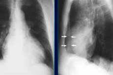
A chest X-ray with fluid accumulation in the pericardial cavity reveals an enlarged silhouette of the heart, but if the volume of effusion is insignificant, the X-ray will not show it.
In chest computed tomography CT signs of hydropericardium are widened cardiac contours with low density (up to 20-30 HU). However, CT and MRI are usually not used to diagnose pericardial effusions, since the most effective imaging method in this case is cardiac ultrasound - echocardiography. And to detect fluid in the pleural cavity - chest ultrasound. [ 9 ], [ 10 ]
Ultrasound signs of hydrothorax and hydropericardium - anechoic (echo-negative) space in the pleural cavity and between the two layers of the pericardium, behind the heart (in the atrioventricular groove). Moreover, in the pericardial cavity, fluid is usually identified only in systole, when the heart moves away from the inner surface of the pericardial sac.
Differential diagnosis
Differential diagnostics are carried out with exudative pericarditis, hemopericardium, and cardiac muscular hypertrophy. Exudative effusion is also differentiated from transudate. [ 11 ]
Who to contact?
Treatment hydropericardium
If possible, treatment of hydropericardium should eliminate its underlying cause, and the choice of method is determined primarily by the etiology. That is, pericarditis or myocarditis, pneumonia or pleurisy, hypothyroidism or cancer are treated. [ 12 ]
In drug therapy of pericardial effusion of inflammatory origin, nonsteroidal anti-inflammatory drugs (NSAIDs) are used, that is, such drugs as: Aspirin (0.7-1 g per day for 10 days); Ibuprofen (0.6 g twice a day); Indomethacin (50 mg twice a day). It should be borne in mind that these drugs are contraindicated in gastritis and stomach ulcers.
For the treatment of hydropericardium caused by microbial infection, antibiotics are prescribed, and in cases of heart failure, diuretics (with monitoring of serum sodium levels).
In case of recurrent effusions, NSAIDs and Colchicine (daily dose - 1 mg) are used, and in cases of systemic inflammatory diseases - glucocorticoids, for example, Prednisolone or Dexamethasone (daily dose is 0.2-0.5 mg per kilogram of body weight). [ 13 ]
You should not use folk remedies on your own – without consulting a doctor – in particular, herbal treatment, taking decoctions of lingonberry leaves, bearberry herb, naked hernia, field horsetail or marsh cudweed. [ 14 ]
Surgical treatment involves removing the fluid that has accumulated in the pericardial cavity, all the details in the publication - Pericardial puncture, pericardiocentesis [ 15 ], [ 16 ], [ 17 ]
If effusion frequently recurs, a minimally invasive procedure may be performed to create a so-called pericardial window, a small opening in the lining of the pericardium to drain the accumulating fluid. [ 18 ]
Prevention
In most cases, there is no way to prevent the occurrence of hydropericardium. [ 19 ]
Forecast
Given that hydropericardium occurs for various reasons, the prognosis for its outcome may not be equally favorable in all cases. Although small accumulations of serous fluid may resolve spontaneously or require minimal therapeutic intervention.