Medical expert of the article

New publications
Gingival recession: causes, symptoms, elimination without surgery, how to stop
Last reviewed: 07.07.2025

All iLive content is medically reviewed or fact checked to ensure as much factual accuracy as possible.
We have strict sourcing guidelines and only link to reputable media sites, academic research institutions and, whenever possible, medically peer reviewed studies. Note that the numbers in parentheses ([1], [2], etc.) are clickable links to these studies.
If you feel that any of our content is inaccurate, out-of-date, or otherwise questionable, please select it and press Ctrl + Enter.
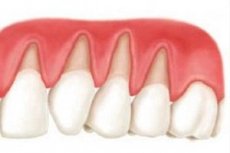
Gum recession (apical displacement of the gingival margin) is a loss of soft tissue of the gum in the vertical direction, which leads to a gradual exposure of the neck of the tooth. According to statistics, this pathological process is more often observed in adulthood, but the tendency for it to occur in children and young people is growing every year. This trend is associated with the high availability of orthodontic treatment with braces, a tendency to chronic stress, urbanization, etc. Often, gum recession does not bother people if it is located on the inner surface of the gum (from the side of the palate). This is explained by the fact that with such localization, the aesthetic properties of a person's smile are not violated, since the defect is not visually determined. However, when even a small loss of gums appears on the side of the lips and cheeks, a person immediately notices it. The tooth seems elongated, which makes it disproportionate to others. And if the teeth are genetically elongated, then recession can create very unpleasant consequences for the aesthetics of the smile.
 [ 1 ]
[ 1 ]
Symptoms gingival recession
Symptoms of gum recession may not appear for a long time. Often, a person is bothered by unpleasant sensations caused by improper placement of a crown or filling, gum inflammation, pain in the teeth, joint, etc. Against the background of these symptoms, the clinical picture of recession looks very poor and unobtrusive. The first signs of this pathological process are the appearance of a small gum defect. Most often, it has the form of a narrow vertical strip. These symptoms indicate that the recession is at the first stage (when the root is exposed to 3 mm). If the process occurs from the mouth, then a person may not pay attention to the presence of a defect. In this case, the recession will not cause any complaints. The second stage is root exposure from 3 to 5 mm. In this case, the defect becomes not only longer, but also wider. At this stage, a person may be bothered by aesthetic defects associated with the loss of gum tissue. Tooth hypersensitivity may develop due to exposure of its cement.
At the third stage, the gum recession is more than 5 mm. At the same time, aesthetic problems become very noticeable, and the symptom of increased tooth sensitivity becomes more pronounced.
In the generalized form, gum recession occurs in the area of 4 or more teeth. If the cause of gum recession was orthodontic treatment with fixed devices, then the complaints will consist of a decrease in the aesthetics of the smile. If the reason for the recession was periodontitis, then a whole range of symptoms will be observed. Inflammation of the gums provokes constant bleeding at the slightest injury. Violation of the gingival attachment leads to the formation of periodontal pockets, from which purulent contents can be released. A person feels pain, itching and tingling in the gum. Saliva becomes viscous, an unpleasant taste appears in the mouth, which does not disappear after brushing the teeth. The gums look bright red, their edges have a swollen, torn appearance, which visually looks very unattractive.
With periodontosis, the gums have a pale pink tint, which indicates the absence of inflammation. However, recession with this disease extends throughout the entire dental row. Progression of gum loss can continue until the entire tooth is completely exposed. Interestingly, significant bone and gum loss does not always lead to significant tooth mobility. This is due to the fact that in the area of the root apex, the process of cement deposition occurs, ultimately leading to hypercementosis. Between the tooth and the remaining gum, a large amount of hard dental deposits (tartar) are deposited, which also prevents excessive tooth mobility.
Forms
Miller's classification of gum recession is the most popular among foreign and domestic specialists. The author divided the types of apical gum displacement into four classes depending on the depth of the lesion.
The first class includes narrow and wide recessions, in which the gingival papillae (triangular areas of the gum between the teeth) and bone tissue are not damaged. The defect does not reach the mucogingival line (the place where the gum passes into the mobile mucosa).
The second class is represented by narrow and wide recessions that reach the mucogingival line and may cross it. The height of the gingival papillae is not changed, the integrity of the bone is not damaged.
Class III recessions may include Class I and Class II recessions, which involve moderate loss of bone or interdental papilla height.
Class IV includes class I and II recessions, in which there is significant loss of bone or interdental papilla height.
In addition to Miller's classification, apical gingival displacement is usually divided by the extent of the defect. If up to three adjacent teeth are involved in the process, then the gingival recession is local. When the defect extends throughout the entire dental row, the pathological process is called generalized gingival recession.
[ 4 ]
Complications and consequences
Recession can be caused by chronic gum trauma. In this case, the local protective forces of the gum tissue are significantly reduced. In this regard, the probability of bacterial flora joining is very high. As a result, gingivitis, localized and generalized periodontitis may develop. However, most often, recession is already a symptom of a certain pathological process. As previously stated, periodontitis and periodontosis are almost always accompanied by a decrease in gum tissue. Exposure of the roots worsens the condition of the teeth and surrounding tissues. As a result of the progression of these diseases, gradual loss or removal of teeth steadily occurs. If periodontitis and periodontosis are not treated, they will continue to exist until the last tooth is removed from the oral cavity. These diseases can create obstacles to rational prosthetics. Any removable and non-removable structures, implants are contraindicated in the presence of inflammatory and destructive processes in the mouth. And recession is one of the reasons that can lead to such conditions.
Diagnostics gingival recession
You can diagnose gum recession even at home. To do this, it is enough to see a soft tissue defect that fits the description in the classification. However, to eliminate the pathological process, it is necessary to understand what caused it. This requires a comprehensive examination in the dentist's office. First of all, anamnesis should be collected. It includes a number of diagnostically important questions:
- When was gum recession first noticed?
- What form did the defect take?
- What complaints are there at the moment?
- When was the last time you had a prosthesis (or filling) done?
- Have you had gum treatment before?
- Was orthodontic treatment performed?
- What kind of brush is used to brush teeth?
- Do you grind your teeth at night?
- Do you have any problems with your temporomandibular joint (pain in the morning, clicking, etc.)
- Do you have any bad habits (biting pencils, nails, etc.)?
Answers to all of the above questions are of great value for diagnosing diseases that have caused gum recession.
The next stage of diagnosing gum tissue loss is an examination of the oral cavity. The dentist determines the condition of the hard tissues of the teeth, fillings, crowns, dentures, the mucous membrane of the gum, tongue, palate, lips and cheeks. Only a thorough and comprehensive examination will reveal the true cause of the defect. Among the instrumental methods, probing is effective. Using a periodontal probe, the doctor evaluates the size of the defect, the integrity of the circular ligament of the tooth (which is completely damaged by periodontitis), the presence and depth of periodontal pockets.
Excellent results in diagnosing recession are shown by the use of various clinical tests. To determine the presence of gum inflammation, the Schiller-Pisarev test is used. The solution of the same name is applied to the gums and the change in the color of the mucous membrane is assessed. If it becomes dark brown, then there is an inflammatory process in the gum tissue. Chronic destructive processes in bone tissue can be indicated by changes in the bone structure on an X-ray, which is also an important diagnostic method.
Among the laboratory tests, clinical blood analysis, blood sugar analysis and general urine analysis are distinguished. These data will help to exclude some systemic diseases that could provoke apical displacement of the gum.
As a result of thorough diagnostics, the recession receives its status. It includes such parameters as the cause of the recession, predisposing factors, depth of the lesion, width of the defect, thickness of the gum, Miller class. These data are a kind of map for effective treatment.
Who to contact?
Treatment gingival recession
Elimination of gum recession is a complex multi-stage intervention that requires precise planning. Before starting treatment, it is necessary to find out what caused the loss of gum tissue. Only after determining the origin of the pathological process will it be possible to understand how to stop gum recession. If this was caused by aggressive tooth brushing, then you should change your toothbrush to a softer one and control the pressure you apply to it. As a rule, recession caused by improper tooth brushing stops getting worse after changing the oral care method. After that, the doctor and the patient decide together whether to correct the gum or leave it as is. If the depth of the lesion is large, then the opinion of the specialist is more significant, since we are talking about the health of the gums of the entire oral cavity. If the recession is insignificant, then the person can decide for himself whether to carry out the intervention. In this case, we are talking only about the aesthetic properties of the gum, so the patient's preferences come to the fore.
If the pathological process is provoked by a poor-quality filling, crown or prosthesis, this indicates that these works are unsuitable. In the near future, the insolvent structures are removed and the fillings are removed. At this stage, a preliminary plan for replacing defects in teeth and dental arches is determined, which will be implemented after gum correction.
In periodontal diseases (periodontitis, periodontosis), pathological processes must be transferred to stable remission. In no case should recession correction be started in the presence of destructive and inflammatory processes in the oral cavity.
Gum correction techniques
There are several methods of closing the recession. According to the classification of H. Erpenstein and R. Borchard, conservative and surgical treatments are distinguished. Surgical interventions are divided into single-layer methods, two-layer methods, targeted tissue regeneration and additional methods.
The conservative method can be used in cases where the cause of recession is aggressive tooth brushing. In this case, the tooth brushing technique is corrected and a periodontal dressing is applied to the damaged area. A special gel with isolating and regenerating properties (for example, GC Coe-Pak) can be used as a dressing. Medication therapy is also recommended. Among the drugs that help close the recession are regenerating drugs (Methyluracil), multivitamin complexes (Aevit, Superia), antiseptics (chlorhexidine, hydrogen peroxide), herbal preparations (infusion of sage, chamomile, rose hips).
Single-layer surgical methods include 5 types of operations. The most popular and simple of them is the coronally positioned flap. The essence of the technique is that a section of soft tissues - a flap - is cut out in the recession area. Then this flap is stretched in such a way as to close the recession. After that, the wound is sutured and the wound heals within several months. Since this method involves using only available tissues, surgical intervention is performed to close small recessions. For the same reason, this method is not used for thin gum biotypes. An important condition is the distance from the recession edge to the mucogingival border, which should be at least 4 mm. If the operation is performed correctly, and the postoperative period passes without deviations, then after two to three months there are no traces of recession and surgical intervention. Other types of single-layer techniques are a laterally displaced flap, a double papillary flap, a crescent flap, and an epithelialized connective tissue graft. All these methods are more complex, demanding of the anatomical conditions of the body and the delicate work of the surgeon. It is very difficult to achieve the simultaneous presence of all factors, therefore these methods are used quite rarely.
The idea behind two-layer techniques is to place a connective tissue graft between the primary flap and the tooth surface. This improves the volume of soft tissue, the regenerative properties of the gum, aesthetics, and the speed of wound healing. The most commonly used flap surgeries are the following:
- Operation by Langer and Langer.
- Operation Bruno.
- Operation Raetzke.
The essence of the Langer and Langer technique is to make three incisions. One incision is horizontal and crosses the recession. Two vertical incisions are located on the sides of the recession, as a result of which the incision line takes the shape of an inverted letter "P". This allows for the separation of a square flap and the placement of the graft between the gum and the tooth.
The Bruno operation is an improved technique of Langer and Langer. The advantages of the updated technique are the absence of vertical incisions. This improves the blood supply to the transplant and the aesthetic properties of the gum in the recession area. However, the protocol of the operation without vertical incisions is more complex.
The Raetzke technique, or "envelope method" can be called the most minimally invasive intervention among the listed two-layer operations. When closing a recession using this method, any vertical and horizontal incisions are excluded. This approach allows preserving the blood supply to the transplant and tissues around the recession. Despite the small volume of surgical manipulations, this technique is quite complex. The surgeon needs to dissect the soft tissues in the defect area and create a so-called "envelope". Since the view of the surgical field is quite limited, this can lead to injury to the underlying tissues. Therefore, all manipulations should be carried out carefully and without haste. After creating a pocket (envelope), the transplant is placed in it, and the wound is sutured.
In addition to the listed flap surgeries, there are many other methods for closing gum recession. The method of guided tissue regeneration is quite popular. In this case, various artificial membranes are used, which are installed instead of a transplant. Although they cannot be compared in effectiveness with a connective tissue transplant, their use is quite popular.
Various nutritional preparations are used as additional means during flap surgeries. For example, gels based on enamel matrix proteins (Emdogain by Straumann) activate tissue regeneration, allowing to increase the probability of the expected result and eliminate recession faster. Also, in combination with single-layer techniques, various allografts and platelet-rich plasma are used. These techniques are currently at the stage of theoretical and practical study, so they are popular mainly in the scientific field.
Laser surgery is currently widespread. It differs from classical surgery only in that a laser is used instead of mechanical cutting instruments (scalpels, scissors). Treatment of gum recession with a laser is not a special and specific operation. The doctor chooses one of the gum correction methods and uses a laser to make incisions. Its advantage is the accuracy of the incisions, the absence of significant bleeding and more active regeneration of soft tissues. However, the laser unit is an expensive device and requires regular maintenance. This entails a higher price for treatment when using it.
Despite the large number of synthetic membranes, regenerating gels and other means, the most effective at present is considered to be connective tissue transplant. Techniques using it allow to close relatively large gum defects with a favorable prognosis for the structure, function and aesthetics of the gum.
In the postoperative period, to speed up the healing of the soft tissues of the gum, it is recommended to undergo a course of physiotherapy. UHF therapy, darsonvalization and fluctuation have a positive effect on regeneration. The course of procedures includes about 10 visits and is adjusted by the attending physician.
Vitamin therapy is one of the components of complex treatment of gum recession. Vitamins of group A, E, C improve the processes of epithelialization and local metabolism, which allows to achieve healing of the wound surface without negative consequences and complications. It is recommended to use complex preparations: for children and adolescents - Pikovit, for children and adults - Superia, etc.
Many homeopathic preparations show high efficiency in maintaining a satisfactory condition of the body in the postoperative period. Such preparations are Lymphomyosot, Traumeel gel, Mucosa compositum, etc. The dosage is indicated by the attending physician, who prescribes a treatment plan. Despite the mistrust of many people, homeopathy has a positive effect when used as an additional therapy.
Medicinal herbs after surgery have an antiseptic, soothing and anti-inflammatory effect. Solutions of chamomile, sage, oak bark and other herbs are recommended for use for 2 weeks after surgery.
After the gum recession has closed, it is recommended to perform rational prosthetics, if necessary. Veneers, metal-ceramic and all-ceramic crowns, bridge prostheses and other orthopedic structures are currently widely used. They will stabilize the load on the teeth and eliminate the appearance of new defects in the soft tissues of the gum.
Many people are fans of folk remedies, homeopathy and herbal medicine. The effectiveness of these remedies in the postoperative period has already been discussed. However, it is worth understanding that with the help of herbs it is impossible to achieve the same result that can be achieved with surgical intervention. Even with small defects, spontaneous closure of the gums is not observed in all cases. There is no guarantee that daily rinsing of the mouth with medicinal solutions will eliminate the loss of gum tissue. Moreover, home treatment can lead to undesirable results. Many drugs inhibit each other's action, accumulate in the body, have a toxic effect on various organs and systems. Therefore, treatment should be agreed with a qualified specialist who can draw up an effective therapeutic plan.
Prevention
Prevention of apical displacement of the gum consists in not allowing the occurrence of predisposing factors for the loss of gum tissue. It is necessary to use a toothbrush of medium or low hardness. See the publication - Hygienic cleaning of teeth - types and features, the procedure for hygienic cleaning of teeth. After installing a filling, crown or prosthesis, you should tell the doctor about all the unpleasant sensations in the mouth after the work is done. Early correction of restorations will eliminate the occurrence of many adverse effects. It is necessary to prevent the occurrence of bad habits and get rid of existing ones. It is important to understand that teeth are intended only for grinding food, and then they will serve you for many years.
Forecast
If the provoking factors are not eliminated, the gum recession will continue to progress and will eventually lead to various complications, which will consist of aggravation of structural, functional and aesthetic deficiencies. If the main causes of recession have been eliminated and high-quality treatment has been carried out, the prognosis is very favorable.