Medical expert of the article
New publications
Gluteal muscles
Last reviewed: 07.07.2025

All iLive content is medically reviewed or fact checked to ensure as much factual accuracy as possible.
We have strict sourcing guidelines and only link to reputable media sites, academic research institutions and, whenever possible, medically peer reviewed studies. Note that the numbers in parentheses ([1], [2], etc.) are clickable links to these studies.
If you feel that any of our content is inaccurate, out-of-date, or otherwise questionable, please select it and press Ctrl + Enter.
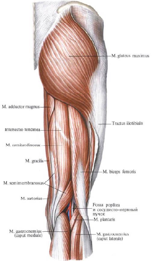
The gluteus maximus (m.gluteus maximus) is strong, has a large-bundle structure, and stands out in relief due to its large mass in the gluteal region (regio glutea). This muscle reaches its greatest development in humans due to upright posture. Situated superficially, it has a wide origin on the ilium (linea glutea posterior), on the initial (tendon) part of the muscle that straightens the spine, on the dorsal surface of the sacrum and coccyx, on the sacrotuberous ligament.
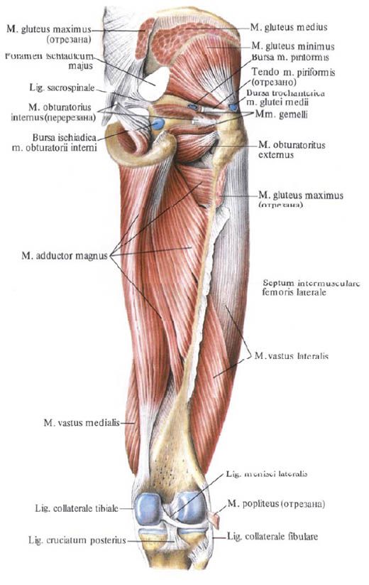
The muscle passes obliquely downwards and laterally and is attached to the gluteal tuberosity of the femur. Part of the muscle bundles passes over the greater trochanter and is woven into the iliotibial tract of the broad fascia. Between the tendon of the muscle and the greater trochanter there is a trochanteric bursa of the gluteus maximus (bursa trochanterica musculi glutei maximi), and at the level of the ischial tuberosity there is a sciatic bursa of the gluteus maximus (bursa ischiadica musculi glutei maximi).

Function: can act on the hip joint with its entire mass or with individual parts. Contracting with its entire mass, the gluteus maximus extends the thigh (simultaneously turning it outward). The anterior superior bundles of the muscle abduct the thigh, tense the iliotibial tract of the broad fascia, and help maintain the knee joint in an extended position. The posterior inferior bundles of the muscle adduct the thigh, simultaneously turning it outward. With a fixed lower limb, the muscle extends the pelvis, and with it the torso, holding it in a vertical position on the heads of the femur (giving the body a military posture).
Innervation: inferior gluteal nerve (LV-SII).
Blood supply: inferior and superior gluteal arteries, medial circumflex femoral artery.
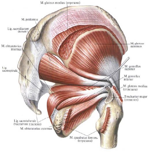
The gluteus medius muscle (m.gluteus medius) originates on the gluteal surface of the ilium, between the anterior and posterior gluteal lines, on the broad fascia. The muscle goes down, passes into a thick tendon, which is attached to the top and outer surface of the greater trochanter.
The posterior bundles of the muscle are located under the gluteus maximus. Between the tendon of the gluteus medius and the greater trochanter is the trochanteric bursa of the gluteus medius (bursa trochanterica musculi glutei medii).

Function: abducts the thigh, the anterior bundles rotate the thigh inward, the posterior bundles rotate it outward. With the lower limb fixed, together with the gluteus minimus, it holds the pelvis and trunk in a vertical position.
Innervation: inferior gluteal nerve (LIV-SI).
Blood supply: inferior gluteal artery, lateral circumflex femoral artery.
The gluteus minimus (m.gluteus minimus) is located under the gluteus medius. It originates on the outer surface of the iliac wing between the anterior and inferior gluteal lines, along the edge of the greater sciatic notch. It is attached to the anterolateral surface of the greater trochanter of the femur; some of the bundles are woven into the capsule of the hip joint. Between the tendon of the muscle and the greater trochanter there is a trochanteric bursa of the gluteus minimus (bursa trochanterica musculi glutei minimi).

Function: abducts the thigh, the anterior bundles participate in the rotation of the thigh inward, and the posterior ones - outward.
Innervation: superior gluteal nerve (LIV-SI).
Blood supply: superior gluteal artery, lateral circumflex femoral artery.
What do need to examine?
How to examine?


 [
[