Medical expert of the article

New publications
Frostbite
Last reviewed: 07.07.2025

All iLive content is medically reviewed or fact checked to ensure as much factual accuracy as possible.
We have strict sourcing guidelines and only link to reputable media sites, academic research institutions and, whenever possible, medically peer reviewed studies. Note that the numbers in parentheses ([1], [2], etc.) are clickable links to these studies.
If you feel that any of our content is inaccurate, out-of-date, or otherwise questionable, please select it and press Ctrl + Enter.
Frostbite is tissue damage caused by local exposure to cold, leading to a prolonged decrease in temperature, damage to anatomical structures, and even to organ necrosis.
ICD-10 code
- X31 Exposure to excessively low natural temperatures.
- T33.0-9 Superficial frostbite.
- T34.0-9 Frostbite with tissue necrosis.
- T35.0-7 Frostbite involving multiple body regions and unspecified frostbite.
Symptoms of frostbite
In the development of pathological changes in the affected areas, the leading role belongs to arterial spasm. With short-term exposure to cold, only superficial vessels react, and frostbite of the 1st and 2nd degree occurs. With more prolonged and intensive cooling, a long-term spasm of all arterial vessels occurs, resulting in the death of soft tissues and bones.
During frostbite, two periods are distinguished: latent (pre-reactive) and reactive, before and after warming the patient, respectively. In the first period, the frostbitten area is pale, cold to the touch, and insensitive. The patient complains of a feeling of numbness, "stiffness," and "cold feet." Less commonly, pain in the feet and calf muscles is a concern. In a small number of observations, frostbite is not accompanied by any sensations. In the pre-reactive period, diagnosis is not difficult, but it is impossible to determine the depth and extent of tissue damage.
In the reactive period after warming up the frostbitten area, the main complaint of patients is pain. It occurs immediately after warming up the patient, is quite intense and is typical for all victims. Patients experience a burning sensation, heat, "stiffness" in the frostbitten areas. Edema and a change in skin color from white to cyanotic indicate the end of the "latent period".
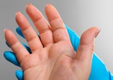
In 95% of cases, frostbite affects the extremities, most often the lower ones; the lesion is limited to the fingers and does not extend above the ankle or wrist joints. Such localization is due to worse blood supply to the peripheral parts of the extremities compared to other areas of the body; they are more susceptible to the effects of cold, and hemodynamic disorders develop in them faster. In addition, the hands and feet are less protected from the effects of cold. Frostbite in other locations (ears, nose, cheeks) is observed much less frequently. In the overwhelming majority of cases, frostbite occurs when exposed to frost at an air temperature of -10 °C and below. However, with high air humidity and strong wind, frostbite is possible at higher temperatures approaching 0 °C. People in an unconscious state (with severe alcohol intoxication, severe trauma, an epileptic seizure) are more often subject to frostbite. In such situations, frostbite of the fourth degree usually occurs.
Atypical forms of frostbite
In contrast to the described “classical” form of frostbite, there are several varieties, characterized by a unique clinical course and arising in conditions different from those described - chills and “trench foot”.
Chilblains are a pathological skin condition that develops as a result of prolonged exposure to low temperatures and high humidity and is characterized by swelling, cyanosis, pain when pressed, and itching. They are considered chronic frostbite of the first degree; eliminating repeated cooling helps eliminate chilblains. Chilblains often occur in the form of dermatitis or dermatoses. In people who, due to the nature of their work, are constantly exposed to cold with high humidity (fishermen, sailors, timber rafters), chilblains are considered an occupational disease.
Trench foot is frostbite of the feet as a result of prolonged moderate cooling; it occurs at an air temperature of about 0 °C and high humidity, mainly in a military situation. This is a form of local cold injury, first described during the First World War in the case of mass lesions of the feet of soldiers who had been in trenches filled with water for a long time. The disease is characterized by disturbances of tactile, temperature and pain sensitivity, the occurrence of pain, and the appearance of a feeling of "woodenness" of the feet. Edema develops, the skin acquires a pale shade with areas of hyperemia, cold to the touch; then blisters with hemorrhagic contents form. The final result is necrosis of the feet with the development of wet gangrene. With bilateral lesions, an extremely severe course of the disease with high fever and severe intoxication is characteristic.
A peculiar form of cold injury is "immersion foot" ("submerged limb"). This pathology develops when limbs are in cold water for a long time and occurs almost exclusively in sailors or pilots in distress at sea with water temperatures from 0 to +10 °C. Two, three, and sometimes four limbs are affected simultaneously, and frostbite occurs 2-3 times faster than on land.
“High altitude foot” occurs in pilots when flying at high altitudes at extremely low air temperatures (from -40 to -55 °C) and high speeds, in conditions of low oxygen content.
Sometimes contact frostbite develops from bare hands coming into contact with metal objects cooled to -40 °C. These frostbites are usually superficial and limited in area.
Complications arising from frostbite are divided into local and general. The most common local complications are lymphangitis, lymphadenitis, thrombophlebitis, erysipelas, phlegmon, abscess, arthritis, and osteomyelitis. Later, neuritis, endarteritis, trophic ulcers, cicatricial deformations and contractures, and persistent increase in cold sensitivity develop. General complications in the early stages include intoxication, pneumonia, sepsis, and multiple organ failure; later, myocardio-, nephro-, and encephalopathy.
Classification
Frostbite is classified according to the depth of tissue damage into 4 degrees:
- Frostbite I. After warming up, the skin of the frostbitten area is bluish, often with a purple tint, slight swelling and marbling of color are possible. Frostbite of the first degree passes after 5-7 days of conservative treatment, with the swelling completely disappearing, the skin acquiring a normal color. Itching, cyanosis, and increased sensitivity to cold remain for a short time.
- Frostbite II. Accompanied by necrosis of the upper zone of the papillary-epithelial layer, formation of blisters filled with transparent serous fluid (sometimes several days after warming). The bottom of the blister is the papillary layer of the skin, represented by a surface of pink or pale red color, sensitive to mechanical irritation. At this degree, the germinal layer of the skin is not damaged, therefore, in a short time (8-14 days), complete epithelialization of the wound surfaces is observed under the influence of conservative treatment. Residual manifestations are similar to those of degree I.
- Frostbite III. The skin of the affected area is deathly pale or bluish-purple! Tissue edema is pronounced. The blisters are filled with hemorrhagic fluid; after opening it and removing the epidermis, the non-viable surface of the papillary layer of the skin is exposed, insensitive to mechanical irritation (for example, a needle prick or touching a ball with alcohol). Necrosis spreads to the entire thickness of the skin. Independent epithelialization of such wounds is impossible due to the death of all epithelial elements of the skin. Healing is possible through the development of granulation and scarring. Lost nails often grow back deformed. Extensive wound defects require plastic closure with autodermal grafts.
- Frostbite IV. Occurs with the longest exposure to a cold agent and a prolonged period of tissue hypothermia, accompanied by necrosis of all tissues, including bones. Dry gangrene of the fingers or toes and wet gangrene of the proximally located areas develops 8-10 days after the injury. The demarcation line appears by the end of the 2nd - beginning of the 3rd week. The process of spontaneous rejection of necrotic tissues takes several months.
In frostbite of grades III-IV, four zones of pathological changes are distinguished (in the direction from the periphery to the center):
- total necrosis;
- irreversible degenerative changes (where trophic ulcers and ulcerative scars may subsequently occur);
- reversible degenerative processes;
- ascending pathological processes.
- In the last two zones, the development of persistent vascular and neurotrophic disorders is possible.
How is frostbite recognized?
The victim indicates a long stay in low air temperature conditions. The differential diagnosis of frostbite is made with gangrene of the toes in diabetic angiopathy or obliterating endarteritis.
Indications for consultation with other specialists
Need consultation with a vascular surgeon and therapist.
Example of diagnosis formulation
Frostbite of both feet, grade III-IV.
What do need to examine?
How to examine?
Who to contact?
Frostbite Treatment
The main goal of treatment is warming and restoration of normal blood flow in the affected parts of the body.
Indications for hospitalization
Frostbite of III-IV degree of any area and location; widespread superficial frostbite.
First aid for frostbite
In order to prevent further cooling and restore the temperature in the affected parts of the body, the victim should be taken to a warm room, changed into dry clothes and shoes. General measures include giving the victim hot tea, coffee, food, 50-100 ml of vodka. In case of frostbite of the ears, cheeks, nose, you can easily rub the frostbitten areas with a clean hand or soft cloth until the skin turns pink.
It is necessary to exclude premature warming from the outside, when the victim is already indoors: the heat should come “from the inside” due to blood circulation. Thus, the boundary of tissue warming gradually shifts to the periphery, where circulation is restored earlier than metabolism, which protects tissues from ischemia. To achieve this effect, a thermal or heat-insulating bandage is applied to the affected area as quickly as possible. It alternates 5-6 layers of gauze and cotton wool (batting, wool, foam rubber, synthetic padding) with two or three layers of compress paper (polyethylene, metal foil) laid between them. The thickness of such a bandage is 5-6 cm. Before applying the bandage, no manipulations are performed with frostbitten areas. Bandages are left on the affected area for at least 6-12 hours, until sensitivity is restored.
After hospitalization of the victim, measures are taken to gradually warm the tissues "from the inside out". This is achieved by infusion systemic and regional treatment, the purpose of which is to eliminate vascular spasm, restore microcirculation, and prevent thrombus formation in small and large diameter vessels.
The use of UV radiation, UHF therapy, infrared irradiation and simply warm air from a fan in the first phase of the wound process in frostbite of grades III-IV helps to convert wet necrosis into dry necrosis.
 [ 10 ]
Drug treatment
To improve blood circulation in the affected limbs, the following drugs are administered intravenously 2 times a day during the first week after the injury: solutions of dextran (rheopolyglucin) 400 ml, 10% glucose - 400 ml, procaine (novocaine) 0.25% - 100 ml, vitamin B: 5% - 2 ml, 1% nicotinic acid - 2 ml, 5% ascorbic acid - 4 ml, drotaverine (no-shpa) 2% - 2 ml, papaverine 2% - 4 ml; sodium heparin (heparin) 10,000 U, pentoxifylline (trenthal) 5 ml or dipyridamole (curantil) 0.5% - 2 ml, hydrocortisone 100 mg. Infusions are performed at a rate of 20-25 drops per minute. Therapy should be continued even if the temperature and tissue trophism have not been normalized within 2-3 days. In this case, it is necessary to reduce the tissue necrosis zone.
Of great importance is the introduction of drugs directly into the regional blood flow of the frostbitten limb. This is achieved by puncturing the appropriate main artery (radial, ulnar, brachial, femoral). The following drugs are usually administered: solutions of procaine (novocaine) 0.5% - 8.0; nicotinic acid 1% - 2.0; sodium heparin (heparin) 10 thousand units; ascorbic acid 5% - 5.0; aminophylline (euphyllin) 2.4% - 5.0; pentoxifylline (trental) 5.0 [or dipyridamole (curantil) 0.5% - 2.0]. On the first day, infusions are performed 2-3 times, in the following 2-3 days - 1-2 times. The duration of the course of vasoactive infusion therapy is at least 7 days.
Novocaine perirenal, vagosympathetic, perineural conduction and simple case blocks performed in the pre-reactive or early reactive periods promote analgesia, vasodilation and reduction of interstitial edema, thereby creating favorable conditions for normalizing the temperature in the affected tissues.
Patients admitted to hospital in the late reactive period, with clearly expressed signs of irreversible tissue damage, should undergo the entire range of treatment and preventive measures described above in order to possibly limit the degree and extent of tissue damage.
Surgical treatment of frostbite
Indications
Deep frostbite of III-IV degree.
Surgical treatment methods
Local treatment of frostbite wounds is carried out according to general surgical rules for treating purulent wounds. It is necessary to take into account the depth of the lesion and the phase of the wound process.
In case of first-degree frostbite, after cleaning the wounds, apply gauze dressings with water-soluble antibacterial creams [chloramphenicol/dioxomethyltetrahydropyrimidine (levomekol), dioxomethyltetrahydropyrimidine/sulfodimethoxine/trimecoine/chloramphenicol (levosin), benzyldimethyl-myristoylaminopropylammonium (miramistin ointment), mafenide], chloramphenicol (syntomycin), etc. Complete epithelialization occurs in a short time (7-10 days) without any cosmetic or functional defects.
In case of frostbite of III-IV degree, conservative treatment allows to prepare the affected areas for surgery. The nature of the drugs used depends on the phase of the wound process. In the first phase (acute inflammation, abundant discharge, rejection of dead tissue), antiseptic solutions, hypertonic solutions of sodium chloride, antibacterial ointments on a water-soluble basis, as well as drugs with a necrolytic effect [trypsin, chymotrypsin, terrilitin, prosubtilin (profezim), etc.] are used. Dressings are done daily, the affected limbs are placed on Beler splints.
In the second phase of the wound healing process (after the inflammation has subsided, swelling and the amount of wound discharge have decreased, and non-viable tissue has been rejected), dressings are changed less frequently (every 2-3 days) with fat-based ointments [with nitrofural (furacilin ointment 0.2%)].
In the third phase (epithelialization and scarring), it is advisable to use biogenic stimulants of plant (Kalanchoe and aloe juice) and animal origin (15% propolis ointment). For the same purpose, ointments with dioxomethyl-tetrahydropyrimidine (methyluracil) 10%, actovegin 20%, etc. are used.
Modern tactics of surgical treatment of deep frostbite pursue the goal of the fastest possible removal of non-viable tissue, prevention of the development of severe complications and maximum preservation of the volume of viable tissue.
As in the treatment of deep burns, necrotomy, necrectomy, amputation and dermatomal free skin grafts are used.
Possible postoperative complications
Suppuration of postoperative wounds, melting of skin grafts, suppuration of donor wounds.
More information of the treatment
Drugs
What is the prognosis for frostbite?
Superficial frostbite has a favorable prognosis, patients return to work. Deep frostbite with damage to large segments of the extremities leads to persistent disability.