Medical expert of the article

New publications
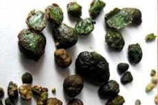
Fecal stones
Last reviewed: 12.07.2025

All iLive content is medically reviewed or fact checked to ensure as much factual accuracy as possible.
We have strict sourcing guidelines and only link to reputable media sites, academic research institutions and, whenever possible, medically peer reviewed studies. Note that the numbers in parentheses ([1], [2], etc.) are clickable links to these studies.
If you feel that any of our content is inaccurate, out-of-date, or otherwise questionable, please select it and press Ctrl + Enter.
Fecal stones are dense formations that form in some cases in the large intestine from its contents. They most often occur in old and senile age. Predisposing factors are long-term stagnation of intestinal contents caused by hypotension or atony of the large intestine, dysfunction of the large intestine in Parkinsonism, congenital anomalies in the form of megacolon, Hirschsprung's disease, additional loops.
Causes fecal stones
Fecal stones occur in old and senile age. Predisposing factors are long-term stagnation of intestinal contents caused by hypotension or atony of the colon, dysfunction of the colon in Parkinsonism, congenital anomalies in the form of megacolon, Hirschsprung's disease, additional loops.
The most important factors contributing to the formation of fecal stones in the colon from its compacted contents are the intensive absorption of water, the slow movement of the contents and the formation of fecal masses. The contents of the small intestine coming from the stomach are liquid, and their passage through the intestine is carried out quickly.
Sometimes a stone that has been closely adjacent to the mucous membrane of the colon wall for a long time becomes “encapsulated,” which helps it to become fixed in this place.
Pathogenesis
Fecal stones can be single or multiple, they are usually round or oval in shape, up to 8-15 cm in diameter. A. Mongo (1830) described an intestinal stone weighing 4 pounds (about 1.9 kg). Intestinal stones have a dense, sometimes very hard consistency, which gave reason to call them stones.
Colon stones consist of compacted fecal matter, sometimes with an admixture of mucus; in some cases, when cut, they have a layered structure (concentric layers are visible). Sometimes fecal stones form around a "core", which can be accidentally swallowed berry seeds that enter the intestine, pieces of meat or chicken bones, unchewed and undigested dense pieces of food, conglomerates formed from poorly digestible dietary fiber, swallowed hair, gallstones, large tablets of poorly soluble drugs, and many other foreign bodies. In some cases, the formation of intestinal stones can be caused by taking large doses of insoluble antacids.
Stones are described which consist almost exclusively of magnesium carbonate, as well as stones which contain 80% of lime carbonate or "fatty-waxy masses", apparently formed from excessive consumption of very fatty foods containing refractory animal fats, or from insufficient digestion of fats.
In some cases, fairly large gallstones enter the intestines through fistulous connections between the gallbladder and the intestines (usually with the transverse colon ), and even urinary stones that enter the intestines through fistulous tracts from the renal pelvis or urinary bladder.
 [ 4 ], [ 5 ], [ 6 ], [ 7 ], [ 8 ], [ 9 ], [ 10 ], [ 11 ], [ 12 ]
Symptoms fecal stones
There may be cramping pain in the abdomen, sometimes ulceration of the intestinal wall, which may cause intestinal bleeding. Large fecal stones may cause intestinal obstruction.
The course of the process during a certain period of time (sometimes very long) is asymptomatic or low-symptom, while in other cases complications arise relatively early.
Where does it hurt?
Complications and consequences
One of the main complications is the occurrence of obstructive (partial or complete) intestinal obstruction. Usually, a spastic component plays a certain role in the development of this complication. The literature describes 6 rare cases of intestinal obstruction when taking large doses of insoluble gel-like antacid drugs. Intestinal bleeding is caused by the formation of bedsores and ulcers of the intestinal wall at the site of adhesion and constant pressure of the intestinal stone. In rare cases, with a long-term existence of the stone and cicatricial-inflammatory changes in the intestinal wall at the site of its adhesion, intestinal stenosis develops over time.
Diagnostics fecal stones
Diagnosis of fecal stones is often difficult. Large stones, especially in the colon, can sometimes be identified using the method of methodical deep palpation. At the same time, compactions along the colon, especially in people suffering from spastic constipation, can often be detected during palpation. If a persistent limited compaction is detected in a patient during abdominal palpation or if a "filling defect" is detected during an X-ray examination of the intestine, a malignant tumor of the intestine should be considered first. If this formation is localized in the colon - cancer, especially since cancerous lesions of the colon are much more common. A number of additional symptoms - mild abdominal pain, loss of appetite, varying degrees of weight loss, mainly old age of patients, accelerated ESR - also suggest a tumor lesion of the intestine, although they can be caused by completely different reasons. Additional examination allows for a more precise diagnosis: plain abdominal radiography and echography allow for the detection of concretions containing calcium salts. If the formation is localized in the large intestine, the correct diagnosis can be made during a rectoscopy or colonoscopy.
What do need to examine?
Differential diagnosis
Differential diagnosis of fecal stones should also be carried out with benign tumors and intestinal polyps.
Who to contact?
Treatment fecal stones
If the diagnosis of "fecal stones" is established, laxatives (in a hospital setting) and siphon enemas (for colon stones) are prescribed for cleansing. If the stone has descended into the rectum, it can be removed with a finger during its examination or, if necessary, with surgical instruments.
If obstructive intestinal obstruction develops, surgery is necessary.