Medical expert of the article

New publications
Colon anomalies
Last reviewed: 07.07.2025

All iLive content is medically reviewed or fact checked to ensure as much factual accuracy as possible.
We have strict sourcing guidelines and only link to reputable media sites, academic research institutions and, whenever possible, medically peer reviewed studies. Note that the numbers in parentheses ([1], [2], etc.) are clickable links to these studies.
If you feel that any of our content is inaccurate, out-of-date, or otherwise questionable, please select it and press Ctrl + Enter.
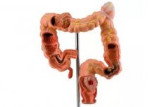
Anomalies of the colon (including the rectum), according to various authors, occur with a frequency of 1:1500-1:5000 newborns. The most common forms of anomalies of the colon are anorectal atresia, atresia of the colon, which are clinically manifested immediately after birth and, as a rule, are incompatible with life.
Sometimes anal stenosis and atresia are combined with fistulas connecting the rectum with the bladder, urethra, and in girls - also with the vagina. Sometimes the fistulous canal goes into the perineum. In some cases, anal stenosis can be widened, and such children survive. In some cases, and with fistulas combined with anal atresia, newborns with this pathology can be provided with surgical assistance.
Congenital megacolon
 [ 9 ], [ 10 ], [ 11 ], [ 12 ], [ 13 ], [ 14 ], [ 15 ]
What causes congenital megacolon?
Congenital megacolon is a hereditary disease, the inheritance pattern is apparently autosomal recessive. Identical twins usually both suffer from this disease. A frequent combination of Hirschsprung's disease with other developmental defects has been noted: with Down's syndrome, enlarged bladder (megacystis), bladder diverticula, hydrocephalus, etc. Congenital megacolon occurs with a frequency of 1:5000 newborns.
Congenital megacolon is a significant expansion of part or all of the large intestine, usually with thickening of the muscular membrane of its wall. Congenital megacolon may be caused by any obstacles to further movement of the contents of the large intestine (stenosis, membranous septa, etc.), but more often it is a congenital defect of its innervation - congenital agacgliosis. This form is usually called Hirschsprung's disease - a congenital absence of nerve ganglia or their insufficient number for normal functioning of the large intestine. In areas of the large intestine deprived of normal innervation, peristalsis is absent or the amplitude and strength of peristaltic waves are significantly reduced, the propulsive ability of the intestine suffers. Most often, these changes are localized in the sigmoid colon (megadolichosigma), but the entire large intestine can be affected (megadolichocolon).
In the pathogenesis of congenital megacolon, aganglionosis of the colon section is of primary importance. As a result, dystrophic changes in the muscular layers of the intestine occur, where the greatest changes in the muscular-intestinal and submucous nerve plexuses occur. Initially, compensatory hypertrophy of the muscular tissue of a more proximal section of the intestine occurs (with regional megacolondolichosigma), later - dystrophic changes in the muscular fibers in these sections (due to constant work overload), as well as their replacement with connective tissue, which leads to an even greater expansion of the intestine proximal to the aperistaltic zone.
Symptoms of congenital megacolon
In most cases, Hirschsprung's disease is detected in the first days after birth - no meconium discharge, abdominal distension, sometimes vomiting. About 20% of newborns develop constant diarrhea due to the development of pseudomembranous colitis. Mortality, despite all measures taken, is very high and reaches 70-75%. Children whose submucosal and muscular-intestinal plexuses are less severely affected and who manage to survive this initial, very difficult period, later suffer from constipation, requiring almost daily enemas and laxatives. Abdominal distension and flatulence are possible. Children suffering from this disease have slow growth, they look younger than their age. They often have anemia, hypoproteinemia, hypovitaminosis.
The diagnosis is confirmed by X-ray examination data (irrigoscopy); in specialized gastroenterological institutions, peristalsis and pressure in the colon are recorded, which also facilitates diagnosis. Particularly indicative is the test with mecholyl (a drug from the cholinomimetic group), after the injection of which the tone and phase activity of normally innervated areas of the colon decrease, but the tone of its denervated areas increases.
Complications of congenital megacolon
In older children and adolescents, due to prolonged retention of fecal matter proximal to the aganglionic zone, gradual and more significant than normal absorption of water and compaction of the intestinal contents, including the ileum, intestinal obstruction may occur. In some rare cases, due to prolonged pressure of fecal matter on the intestinal wall, so-called fecal ulcers occur, and sometimes perforation of the intestinal wall with subsequent development of peritonitis.
Differential diagnosis of Hirschsprung's disease in adults is carried out with organic obstruction of the colon - stenosis, compression, including adhesions after operations performed in the past for another reason, narrowing or obstruction of the intestinal lumen by a tumor, a large polyp, etc. In some regions of South America, a disease caused by Trypanosoma of Cruc (Chagas disease) is quite common, in which the nerve plexuses of the digestive tract are affected. Subsequently, people who have had an acute form of this disease develop megacolon, megaesophagus, gastric dilation, atony and dilation of the gallbladder and urinary bladder; moreover, the changes are most often localized in the esophagus (creating difficulties in differential diagnosis with achalasia of the cardia ) or in the colon (differential diagnosis with congenital megacolon).
Additional loops of the sigmoid colon
Additional loops of the sigmoid colon with some expansion and almost constant constipation are also possible. In some cases, over many years, prolonged stool retention for several days can be of a purely neurogenic "psychogenic" nature; it also occurs in Parkinsonism, long-term use of a number of drugs that weaken the tone and peristalsis of the digestive tract, etc. Naturally, due to the accumulation of feces, the colon, especially in the distal sections, will expand. With systemic scleroderma and some other diseases, atrophy of the muscular layers of the intestinal wall occurs, the tone of the colon decreases, and it expands. However, with systemic scleroderma, there are a number of external signs characteristic of this disease (acrocyanosis, Raynaud's syndrome, trophic ulcers on the tips of the fingers and toes, the "purse-string" symptom - tightening of the skin into folds around the mouth, etc.), which in most cases allow a diagnosis to be made.
In relatively small areas of colon aganglionosis, surgical treatment is possible, and several modifications of the operations have been proposed. In other, usually milder cases, siphon enemas with a large volume of liquid - up to 2 liters - must be administered throughout life, Vaseline oil must be taken orally to improve the passage of the contents of the colon, and therapeutic exercises specially selected for this disease must be performed.
[ 19 ]
Congenital diverticula of the colon
Other anomalies of the colon include congenital diverticula, duplication of the colon and small intestine, communicating or not communicating with the intestinal lumen; the latter resemble cyst-like cavities. Diagnosis is difficult. They are either asymptomatic for a long time or manifest themselves as pain caused by overstretching of the cyst by the contents or intestinal obstruction. Contrast X-ray examination sometimes reveals duplication of the intestine communicating with its main tract. Perhaps ultrasound examination and computed tomography facilitate the diagnosis of this rare anomaly. In most cases, diagnosis is possible during laparotomy. Treatment is surgical.
What do need to examine?
Who to contact?