Medical expert of the article

New publications
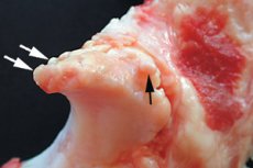
Edge osteophytes
Last reviewed: 29.06.2025

All iLive content is medically reviewed or fact checked to ensure as much factual accuracy as possible.
We have strict sourcing guidelines and only link to reputable media sites, academic research institutions and, whenever possible, medically peer reviewed studies. Note that the numbers in parentheses ([1], [2], etc.) are clickable links to these studies.
If you feel that any of our content is inaccurate, out-of-date, or otherwise questionable, please select it and press Ctrl + Enter.
There are many types of bone growths. If such growths are formed on the end parts as a marginal overgrowth due to deforming loads or a disorder of calcium metabolism, they are called "marginal osteophytes". The problem can be asymptomatic, but most often there is a limitation of mobility and pain in the affected joint. In general, marginal osteophytes are a specific radiological indicator of degenerative processes, their appearance is associated with the development of involutional changes in bone tissue. [1]
Epidemiology
The most common cause of the formation of marginal osteophytes is considered to be osteoarthritis. Among the most frequent manifestations of pathology are pain in the affected joint, morning stiffness. As you move, the pain may decrease somewhat, but by evening it increases again, which is associated with the load.
The involvement of genetic predisposition is not uncommon. On average, symptoms appear between the ages of 40 and 50. Men are more prone to early onset of symptoms. In women, the signs of marginal osteophytes are detected somewhat later, but they are more pronounced - in particular, the pain syndrome is brighter and more intense. The problem more often makes itself known with the onset of menopause.
Causes of the marginal osteophytes
The most common cause of the formation of marginal osteophytes are disorders of metabolic processes. Often the growths arise due to increased loads on a particular joint, which entails damage to the cartilage. Other probable causes include direct trauma to the joint or spinal column. [2]
Specialists point to such main causes of pathology:
- Inflammatory changes in the bone tissue;
- Degenerative changes;
- Bone fractures;
- Prolonged forced stay in one position;
- Tumor processes;
- Endocrine disruption.
Among inflammatory pathologies, the most common is osteomyelitis, a disease in which all bone components are affected, from the periosteum to the bone marrow. Inflammation is provoked by festering bacterial flora or mycobacterium tuberculosis. The primary causes of osteomyelitis are: open bone fractures, foci of chronic infection, violation of recommendations for the safe management of osteosynthesis operations. The disease more often affects the femur, humerus, tibia, upper and lower jaw.
Degenerative intraosseous processes develop against the background of age-related changes in tissues, excessive loads on the articular area. "The culprits" can be deforming spondylosis or oseoarthrosis.
Often, marginal osteophytes are formed after the integrity of the central segment of the bone has been compromised. In the area of the fracture, a specific bone connective tissue callus is formed over time, which is subsequently replaced by osteoid tissue. In the course of regeneration in the circle of displaced bone elements and the tissue of the callus, osteophytes, referred to as posttraumatic, arise. Sometimes outgrowths are formed from the periosteum, which after detachment ossifies and degenerates into a bone formation. Such a phenomenon is not uncommon for injuries to the elbow or knee articulation. Osteophytes can also be caused by tears of ligaments and joint bursae.
Prolonged stay in an uncomfortable, forced position almost always overloads one or another joint, which leads to changes and destruction of cartilage tissue and then bone, which begins to grow with the formation of marginal osteophytes. In addition, the risk of developing deforming spondylosis and osteoarthritis increases.
Sometimes osteophytes grow when the bone is affected by a benign or malignant neoplasm, or as a result of metastases from other structures settling in the bone. This most commonly occurs in patients with osteogenic sarcoma, osteochondroma, ewing's sarcoma, breast cancer or prostate.
As for endocrine pathologies, most often the growth of osteophytes is provoked by acromegaly, a disease accompanied by increased synthesis of growth hormone. The disorder is caused by the formation of a benign mass in the anterior lobe of the pituitary gland.
Vertebral osteophytes arise as a result of deforming spondylosis. In this disorder, the growths appear from the anterior edge of the vertebral bodies, or come from the articular processes.
Risk factors
Regular loads on joints, including the spine, over time cause degeneration of joint surfaces and intervertebral discs, as well as their wear and tear. If such factors as age-related changes, traumatic injuries, bone curvatures are combined, then the unfavorable effect on bone structures and joints increases significantly. The ligamentous apparatus suffers: ligaments thicken, calcium salts accumulate in them. Increased joint friction accelerates the growth of osteophytes.
Degeneration processes in tissues start at a young age, although such changes are gradual and do not become apparent until about 50 years of age. However, there are known factors that can accelerate this process:
- Congenital, hereditary anomalies, deformities;
- Dietary habits (this can also include obesity);
- Peculiarities of lifestyle (hypodynamia, incorrect posture, forced frequent incorrect body position, etc.);
- Injuries (whether sports, domestic or occupational).
Pathogenesis
The formation of marginal osteophytes begins with dysregulation of chondrogenesis involving differentiation of chondrogenic cells located in the periosteum, resulting in the formation of a cartilage-like structure called a chondrophyte. The chondrophyte then undergoes ossification to form a chondroosteophyte, and the entire structure eventually transforms into bone to form an osteophyte. [3], [4]
Although marginal osteophytes have been identified as a sensitive and early sign of cartilage lesions in patients with osteoarthritis, the exact pathogenesis of osteophytes is only beginning to be understood. The cytomorphologic findings and gene expression patterns during osteophyte formation resemble those of fracture bone callus healing and endochondral growth plate ossification. [5] It has recently been shown that osteophyte formation and the presence of cartilage lesions are physically independent phenomena. [6], [7] Previously published studies have shown that osteophyte growth is due to the release of cytokines from damaged cartilage rather than mechanical actions on the joint capsule, that synovial tissue plays an important role in the regulation of osteophyte formation, and that exogenously administered cytokines can induce or inhibit osteophyte formation. [8]
Edge osteophytes are often formed after moderate to severe traumatic injuries, bone fractures, degenerative-dystrophic changes involving joints and the spinal column. The involvement of an inflammatory reaction involving bone or surrounding tissue is not uncommon.
In general, an osteophyte is a pathologic outgrowth of bone tissue. The term is related to the Greek words osteon - bone and phyton - spur, plant. Outgrowths can be single or numerous, different in configuration (thin spikes, serrated formations, tubercles). The structure of osteophytes does not differ from the structure of normal bone tissue.
There are growths like this:
- Bone compact;
- Bone-spongy;
- Bone and cartilage;
- Metaplastic.
Bone-compact osteophytes are made of the compact substance of the bone. It is very strong and can withstand intense physical stress, and is essentially the outer layer of the bone. In addition, the compact substance accumulates certain chemical elements, including phosphorus and calcium. This bone layer is characterized by homogeneity and is present in large quantities in the middle segment of tubular bones.
Bone-compact osteophytes are most often found on metatarsal bones, finger phalanges, and end segments of tubular bones.
Bone spongy osteophytes are formed from spongy tissue, which has a cellular structure and is formed from plates and trabeculae. This substance is light and not particularly strong, it is present in the end segments - epiphyses - of tubular bones and fills almost the entire volume of the spongy structures.
Bony spongiform osteophytes develop under the influence of overload in any part of the spongy or tubular bones.
Bone and cartilage osteophytes appear in cartilage distortions, which may be caused by mechanical overload, inflammatory or degenerative processes in the joint, in which the cartilage tissue thins and undergoes destructive changes. Such marginal growths are most often found in large joints that are subjected to maximum load (e.g., the hip joint).
Metaplastic marginal osteophytes are formed when one cell type is replaced by another cell type. Bone tissue is represented by osteoblasts, osteocytes and osteoclasts. Young matrix-producing structures are osteoblasts, which later transform into osteocytes that lose the ability to divide and produce intercellular matrix. Osteocytes take part in metabolic processes, keep the constancy of organic and mineral composition. As for osteoclasts, their formation is associated with leukocytes, and their main function is the destruction of old bone tissue.
The appearance of metaplastic marginal osteophytes is caused by inflammatory or infectious processes in the bone tissue, or a violation of its regeneration.
Osteophytes in the spine can be classified not only by their structure, but also by location. Thus, experts distinguish:
- Anterior or posterior osteophytes;
- Anterolateral marginal osteophytes;
- Posterolateral osteophytes (especially dangerous when they are formed in the neck area, due to their unfavorable effect on the spinal cord).
Marginal osteophytes of the closure plates are a consequence of degenerative-dystrophic pathology of the spinal column. They arise as a result of compaction of the structure in the intervertebral space (in the upper and lower parts of the intervertebral discs). The problem manifests itself with pronounced neurological symptomatology.
Symptoms of the marginal osteophytes
The most typical symptoms of marginal osteophytes are:
- Pain in the affected joint (dull, pressing, stabbing);
- Limitation of motor capabilities of the affected limb or back (develops gradually, slowly increasing);
- The curvature of the joint;
- Soft tissue swelling.
At the early stage of osteophyte formation, the patient does not feel pain. Sometimes it is just a slight discomfort, in which the patient does not hurry to consult with doctors. Medical help is usually resorted to only with the development of an intense degenerative process, destruction of cartilage tissue, the appearance of a pronounced clinical picture. Patients complain of sharp or aching pain, especially intense against the background of physical activity. If the anterior marginal osteophytes of vertebral bodies are affected, pain in the spine may be felt even when coughing or sneezing. [9]
Painful sensations tend to irradiate, i.e., they radiate to nearby organs and joints, which significantly complicates diagnosis. Edge osteophytes of vertebral bodies may additionally cause such nonspecific symptoms as headache, dizziness, visual and auditory disorders, and so on. The appearance of such signs is caused by the compression of the vascular network supplied by the growths.
Large marginal osteophytes of the articular surfaces lead to significant impairment of joint mobility, which is associated with blockage of movement by the formed growths. The joint capsule thickens, contractures develop: the patient gradually loses the ability to move adequately. In advanced cases, there is a complete destruction of cartilage tissue.
Edge osteophytes of the knee joint are also initially manifested by slight discomfort. Over time, the sensations become increasingly painful and unpleasant. Additional signs include:
- Swelling in the knee;
- Gait disturbances, limping.
Similar symptoms are found if marginal osteophytes of the ankle joint or femur occur.
The main symptom that accompanies marginal osteophytes of the lumbar vertebrae is pain that does not respond well to the use of conventional analgesics. Over time, the mobility of the lumbar region is limited, the patient becomes difficult to turn the body to the side, bend. In severe cases, urination may be impaired. [10]
Osteophytes marginal thoracic osteophytes are accompanied by such pathologic symptoms:
- Pain between the shoulder blades, sometimes radiating to the scapula, arm, shoulder;
- Increased pain syndrome with deep breathing, coughing or sneezing;
- Increasing weakness of the arm on the affected side.
The femoral condyles can be affected by a direct fall on the knee or a strong blow to it. Edge osteophytes of the condyles are accompanied by pain in the knee joint, which requires a distinctive diagnosis with injuries, fractures. In most cases, radiography is sufficient.
Edge osteophytes of the patella make themselves known by pain and crunching in the knee area. The intensity of symptoms is individual: the number and size of the growths play a role. Large bony growths significantly increase the risk of meniscus and ligament damage.
Edge osteophytes of the hip joint can create difficulties in freedom of movement, making it difficult to perform simple activities such as lifting the leg, walking or sitting for long periods of time. Some patients indicate the appearance of stiffness, the feeling that the affected leg does not "obey" them. Possible pain in the buttocks, thighs, lower back.
Marginal osteophytes of the roof of the acetabulum are accompanied by these signs:
- Pain in the thigh, groin area (especially in the morning or after physical activity);
- Stiffness, stiffness;
- Pain when attempting to rotate the lower extremities;
- Limp;
- Crunching;
- Muscle and lumbar pain;
- Inability to walk long distances.
Marginal osteophyte of the tibia reveals itself by the appearance of dull, aching pain in the area of projection of the pathological focus, with intensification after physical activity, during loading, turning. Weakness of the corresponding muscle groups, rapid fatigability, numbness and tingling, swelling of soft tissues are also characteristic.
Edge osteophytes of the shoulder joint show these nonspecific signs:
- Pain with exercise;
- Crunching in the affected shoulder;
- Aching pain at rest;
- Impaired mobility of the shoulder, restriction of some movements.
Edge osteophytes of interphalangeal joints are manifested by pain, burning, tingling, numbness in the area of lateral surfaces of distal and dorsal-lateral surfaces of proximal interphalangeal joints. At the same time there may be stiffness, reduction in the motor volume of the affected joints. Deformity of the affected hand is possible with pronounced growths.
Complications and consequences
Edge osteophytes of the cervical region can provoke the development of vascular disorders, severe headaches, dizziness, ringing and tinnitus, visual disturbances, blood pressure fluctuations. As a result of enlargement of the growths there is a narrowing of the spinal canal, arterial trunks and nerves are pinched, spinal stenosis appears. [11] There is a symptom of "false claudication": the patient feels persistent pain, the lower limbs are numb and "disobey". The discomfort does not disappear even at rest.
Subchondral sclerosis and marginal osteophytes often cause the formation of intervertebral hernia, which in turn provokes the appearance of pain and dysfunction in various organs, numbness of the extremities.
The main unfavorable consequences are associated with the constant growth of marginal osteophytes. The gradual increase in the growths entails compression and displacement of tissues, mechanical damage to nearby structures. In the absence of treatment, the affected joint can completely lose its function, the patient becomes disabled.
To prevent the development of complications, you should seek help from specialists already at the stage of initial symptoms. A specialized doctor will evaluate the visible pathological signs, conduct an examination, and diagnose the problem using a comprehensive examination.
Diagnostics of the marginal osteophytes
Diagnostic measures begin with a direct clinical examination. A medical specialist carefully examines the patient, conducts a neurological examination, assesses the function of nerve endings, identifies their likely compression. Based on a detailed examination, studying the patient's medical history and complaints, the doctor determines further diagnostic tactics.
Particular attention is drawn to such signs:
- Joint pain on movement and at rest, after physical activity and regardless of it;
- Joint curvature, axial deformities;
- Limitation of motor activity, inability to perform active or passive movements.
Lab tests:
- Synovial fluid study;
- Evaluation of biomolecular markers in serum, joint fluid, liquor, etc.
Instrumental diagnosis is usually represented by the following procedures:
- Radiography (allows to detect narrowing of the joint gap, areas of subchondral osteosclerosis, directly marginal osteophytes and signs of subchondral osteoporosis).
- Arthroscopy (visualizes intra-articular structures, allows biopsy).
- Arthrosonography (ultrasound joint examination).
- Computed tomography (layer-by-layer visualization of the joint).
- Magnetic resonance imaging (an informative procedure that does not carry radiation exposure).
- Histomorphologic examination (tissue biopsy).
Diagnostic measures should be carried out comprehensively, using an individualized approach to patients.
Differential diagnosis
Overgrowth of marginal osteophytes should be distinguished from such pathologies:
- Acute arthritis;
- Injuries (meniscus or ligament tear with hemarthrosis, fractures);
- Infectious pathologies, microcrystalline arthritis and other inflammatory intra-articular processes, hemophilia;
- Viral infectious diseases, osteoatrosis;
- Cancer, osteochondroma;
- Gout;
- Other arthritis, arthrosis, arthropathies;
- Herniated discs.
For differential diagnosis, in most cases, regtgenography is sufficient. Sometimes computerized or magnetic resonance imaging is additionally prescribed.
Who to contact?
Treatment of the marginal osteophytes
Treatment of marginal osteophytes begins with the impact on the underlying disease. The standard therapy regimen includes the following methods:
- Conservative treatment (elimination of inflammation and pain syndrome, restoration of local metabolism, tissue repair with non-steroidal anti-inflammatory drugs, chondroprotectors);
- Physiotherapy (at the doctor's discretion);
- Physical therapy (helps relieve muscle spasms, improve metabolism, redistribute the load on the joints);
- Massage;
- Lifestyle correction (eradication of bad habits, development of work and rest regimen, development of stress resistance, elimination of hypodynamia);
- Use of supportive and protective devices, orthoses, corsets, inserts, etc., as indicated;
- Nutritional correction (refusal of unhealthy food, expanding the diet with plant foods and dishes rich in calcium and magnesium);
- Weight normalization.
These therapeutic methods will not eliminate the existing marginal osteophytes, but they can stop further progression of the pathology and relieve symptoms. Surgical intervention is performed to completely remove osteophytes.
To alleviate the patient's well-being, such medications are prescribed:
- Non-steroidal anti-inflammatory drugs (Diclofenac, Ibuprofen, Ketorol, etc. In the form of tablets, capsules, ointments, injections) to eliminate pain and inflammation;
- Corticosteroid drugs (in case of severe pain syndrome, it is possible to inject them directly into the joint cavity);
- Other analgesics, antispasmodics (Midocalm).
It should be understood that all the above medications allow only to alleviate the patient's well-being. However, they cannot eliminate marginal osteophytes.
A certain role in the restoration of the joint structure is played by chondroprotectors: chondroitin, glucosamine and analogs. Such drugs allow you to saturate the tissues of the joint with nutrients, stop the process of degeneration, start cell renewal. True, chondroprotectors are effective only at the early and middle stages of osteophyte development, and also require systematic and prolonged intake. To enhance the action of chondroprotectors, other drugs that can optimize tissue microcirculation are also used. To slow down the processes of cartilage destruction, antienzyme agents are used.
As adjunctive therapy prescribed:
- Physiotherapy (shockwave treatment, automated electromyostimulation, ultraphonophoresis, ozone therapy);
- Physical therapy;
- Exercise LFK (mechanotherapy);
- Joint traction to reduce stress on the affected joint;
- Chiropractic care.
In severe advanced cases, the only effective method of treatment is surgery - corrective osteotomy, involving the removal of part of the bone with the growth, or endoprosthesis - replacement of the affected joint with a prosthesis.
Prevention
Dosed regular physical activity is important to prevent the formation of marginal osteophytes. Adequate sports training, daily gymnastic exercises can improve periarticular blood circulation and optimize tissue nutrition. It is recommended to systematically engage in swimming, dancing, aerobics, take daily walks.
Body weight control is a prerequisite for successful prevention. Excess weight is a direct pathway to musculoskeletal diseases, including the development of marginal osteophytes.
In addition, you should not lift and carry too heavy objects, in any way overload the joints and spine. Do not forget about a full and varied diet, enriched with vitamins and minerals. Among the particularly useful products: greens, vegetables, milk and cottage cheese, hard cheeses, seafood.
Water balance is equally important. Doctors recommend drinking plain, clean water a little at a time throughout the day.
It is necessary to give up all known bad habits. It is proven that smoking, as well as alcohol abuse or drug addiction, have an extremely negative impact on the condition of the bone and cartilage system.
Uncomfortable clothes and shoes, high heels can gradually provoke changes in the joints. Not only the foot area can be affected, but also other joints of the musculoskeletal mechanism.
Forecast
The outcome of the disease depends on its form, degree, and the timeliness and quality of treatment measures. Edge osteophytes often become the cause of disability. Neglected cases are accompanied by the loss of the ability to move and serve themselves. With significant osteophytes of the knee and/or hip joints, the patient may be assigned the first or second group of disability, which depends on the stage of the pathological process and the extent of the lesion.
Edge osteophytes progress slowly enough. If you contact doctors in the early stages of the disease, it is often possible to practically stop the further formation of growths and preserve the motor capabilities of the joints. In the absence of treatment, the risk of irreversible changes in the affected joint increases dramatically.