Medical expert of the article

New publications
Osteophytes of the lumbar spine
Last reviewed: 29.06.2025

All iLive content is medically reviewed or fact checked to ensure as much factual accuracy as possible.
We have strict sourcing guidelines and only link to reputable media sites, academic research institutions and, whenever possible, medically peer reviewed studies. Note that the numbers in parentheses ([1], [2], etc.) are clickable links to these studies.
If you feel that any of our content is inaccurate, out-of-date, or otherwise questionable, please select it and press Ctrl + Enter.
Severe low back pain can indicate the onset of serious problems with the spinal column - in particular, lumbar osteophytes that develop in patients with spondylosis are often the cause of pain.Vertebral body osteophyte is a common form of osteoarthritis, defined as an abnormal bone growth or bone spur that forms along the intervertebral joints. [1] In the later stages of osteophyte development, neighboring vertebrae fuse together to form a bony bridge over the intervertebral disc, called a bridging osteophyte.
If untreated, the pathology can cause the development of a number of complications, up to disability. To prevent this, it is necessary to consult doctors in a timely manner and use all possible ways to solve the problem - from conservative therapy, physiotherapy and physical therapy to surgical treatment, which is indicated in advanced cases. [2]
Epidemiology
Osteophytes are often the result of age-related changes in the spine. Over the years, the bones and ligaments of the spinal column wear out, bone overgrowths are formed. In addition, there is degeneration of intervertebral discs, they weaken, there are protrusions and hernias. Heavy physical exertion (including that associated with professional sports) worsens the situation.
Lumbar osteophytes are somewhat less common than cervical osteophytes. The first symptoms are more often detected in people over 50 years of age, although sometimes it happens much earlier - in 40 and even in 20 years. The rate of growth formation depends on both hereditary factors and the frequency of trauma and stress on the spine. Significant osteophytes can be found in 20-25% of vertebrae aged 20-45 years and in 73-90% of vertebrae over 60 years of age [3]
Men suffer from osteophytes more often than women. This is most likely due to the peculiarities of men's professions and lifestyle. In addition, the disease is characteristic of people who lead inactive lifestyles, such as office workers, truck drivers and others. [4]
Causes of the osteophytes of the lumbar spine
Osteophytes of the lumbar spine are bony growths directly on the vertebrae or their articular processes. They have the appearance of rather sharp protrusions, spikes, etc. Such growths appear for different reasons and differ in their clinical picture. Pathology associated with the formation of osteophytes is called spondylosis.
Depending on the cause of appearance, osteophytes are:
- Post-traumatic;
- Degenerative (dystrophic);
- Marginal (massive);
- Periosteal;
- Neurogenically conditioned.
Post-traumatic osteophytes of the lumbar region arise due to damage to the bone structure. In the spine, such growths are not found as often as in the joints.
Dystrophic osteophytes are due to osteoarthritis or deforming spondylosis.
Marginal (massive) osteophytes of the lumbar region develop with metastasis of malignant processes from the prostate or breast, bone cancer.
Periosteal osteophytes form from the periosteum as a result of long-term inflammatory reactions.
Neurogenic osteophytes are associated with psychological disorders, nervous breakdowns, psycho-emotional shocks.
In addition, the appearance of lumbar osteophytes may have an association with systemic skeletal changes. [5]
Risk factors
The most common factor in the appearance of osteophytes of the lumbar spine is age-related changes in it (structural modifications, mineral accumulations). This process is stimulated by hypodynamia, predominantly sedentary lifestyle, improper nutrition, unfavorable ecology, and bad habits.
Some of the major provoking factors include:
- Hereditary predisposition (if close relatives have been diagnosed with spondylosis, the risk of osteophyte formation increases significantly, even regardless of age).
- Abnormalities of the spinal column (displacement and curvature of the discs of the lumbar spine, and the associated friction of the vertebrae against each other).
- Traumatic injuries to the back and spine.
- Metabolic disorders (disorder of calcium metabolism).
- Infectious and inflammatory processes in the spine.
- Prolonged physical exertion, overloading involving increased fragility or wear and tear of the lumbar vertebrae.
- Overweight, rapid weight gain.
- Endocrine Disorders.
- Neurological diseases.
- Lumbar spine curvature, flat feet.
Pathogenesis
In a healthy state, the vertebrae are connected with the help of discs, which are a kind of shock absorbers that ensure the mobility and flexibility of the spinal column. With the development of degenerative processes, the space between the bony elements narrows, the structure of the edges changes and protrusions or outgrowths - osteophytes - are formed on them. Depending on the location, osteophytes of the lumbar spine are:
- With the rear ones;
- Anterolateral;
- With the front ones;
- Posterolateral.
Anterior lumbar osteophytes grow on the anterior portions of the vertebral bodies. They more often affect the thoracic region, but can also be found in the lumbar part of the column.
Beaked osteophytes of the lumbar spine are anterolateral growths. They are called so because they have an unusual shape in the form of a bird's beak.
Posterior lumbar vertebral osteophytes occur on the posterior vertebral surfaces, often accompanied by pain due to compression of the nerve trunks of the intervertebral foramen.
Posterolateral growths are dangerous in creating compression of spinal structures, but are relatively rare in the lumbar region.
In most cases, single osteophytes in the form of spikes are found. Multiple and more massive growths are less common.
The pathogenetic process proceeds through the following steps:
- The bone tissue increases in volume;
- The disk or ligaments ossify.
Under the influence of various provoking factors, intervertebral discs undergo changes in biochemical processes, which subsequently causes a decrease in the level of moisture and proteoglycans in them.
The development of spondylosis is conventionally considered the final stage of osteochondrosis, so the pathology is more common in the elderly and those who follow a sedentary lifestyle. Due to age or dystrophic changes, collagen fibers that form the fibrous sheath are destroyed. This leads to a deterioration of the shock-absorbing ability of the intervertebral discs. Ligaments lose tone and become brittle. The vertebrae begin to press on the intervertebral discs, as a result of which they flatten.
As a result of these processes, the roots of spinal nerves are affected, which causes the appearance of neurological picture. Increased load on the vertebrae, loss of shock absorption leads to bone overgrowth, which is a kind of compensatory reaction: the bone adapts to new conditions through the formation of outgrowths. Osteophytes can be different in configuration and size, sometimes they grow and seem to "ring" the disk.
If not treated promptly, osteophytes of the lumbar spine can fuse (fuse), which leads to the vertebrae growing against each other. As a result, the mobility of the lumbar spine is blocked, blood circulation is impaired, vessels and tendons are affected, and severe neurological symptoms occur. [6]
Symptoms of the osteophytes of the lumbar spine
The course of the pathology has three clinical stages:
- Osteophytes do not leave the vertebrae, so there is little or no symptomatology.
- The growths extend beyond the vertebrae, which causes periodic pain syndrome - particularly after physical activity.
- The outgrowths become large, uniting two or more vertebrae, which is manifested by impaired mobility and causes pronounced flesh tension.
The symptoms of lumbar osteophytes include, first of all, localized pain in the lumbar region. If nerve compression occurs, the pain radiates to the extremities - in particular, with lumbar compression, the pain gradually descends to one of the lower extremities and the foot.
Pain syndrome may increase with prolonged standing or sitting, leaning to the front. There may be numbness, tingling, weakness in the limb.
The reasons for patients to go to doctors in most cases are pain, as well as:
- Weakness in one or both limbs;
- Bowel or bladder disorders;
- Loss of sensation in the groin area.
Weakness in the upper extremities is also noted less frequently.
If you seek medical help in a timely manner, in most cases it is possible to stop the development of osteophytes without surgical intervention.
In the lumbar spine, there is always a maximum load, relative to other parts of the spine. Therefore, when osteophytes form here, there is a vivid clinical picture. Patients complain of pain in the lower back, with an increase in pain when staying in an uncomfortable position for a long time or prolonged standing/sitting.
Often, marginal osteophytes of the lumbar vertebral bodies create a kind of "obstacle" for turning the body. Thus, it becomes impossible to complete the movement of the torso. Pain appears when the growth irritates a muscle, tendon or nerve bundle, or compresses the spinal cord.
Pain syndrome is aggravated by staying in the same or uncomfortable position for a long time, as well as simply by physical activity. In the late stages of osteophyte development, a pronounced neurological picture is revealed, reflexes are reduced, and the limb musculature atrophies. [7]
Complications and consequences
Progression of osteophyte growth in the lumbar spine can lead to a number of complications. The most serious of these is considered to be radicular syndrome, or lumbar radiculopathy - a pathology caused by compression of one of the roots L1-S1. The complication is characterized by severe lumbar pain, "recoiling" in the leg, numbness, paresis, muscle weakness. In particularly severe cases, paralysis of the limb and pelvic organ dysfunction develop.
Previous studies [8], [9] have shown that vertebral osteophytes cause significant changes in the resistance and flexibility of the functional units of the spine under quasi-static or physiologic loading conditions. Significant vertebral osteophytes increase the stiffness and load-bearing capacity of spinal segments. They also affect the nature, location, and prognosis of vertebral fracture risk. [10]
Osteophytes of the lumbar spine can cause curvature of the spine in the corresponding area, as well as a significant limitation of motor activity. Often patients lose the ability to bend to the side or to the front, put on shoes and tie shoelaces.
Osteophytes of the lumbar region develop quite slowly, but if you delay visiting a doctor, the consequences of the disease can be quite serious. First of all, it is advisable to consult such doctors as an orthopedist or a vertebrologist. After carrying out the necessary diagnostic measures, it may be necessary to consult a neurologist. During the rehabilitation period, a physiotherapist and a specialist in physical therapy are included in the treatment.
Among the most common complications of spinal osteophytes are:
- The formation of bone spurs;
- The development of osteoarthritis;
- Deterioration of motor capabilities up to the point of disability.
Diagnostics of the osteophytes of the lumbar spine
Diagnostic measures for suspected lumbar osteophytes may include these procedures:
- Radiographs;
- Multiple projection CT scans;
- MRI;
- Electroneuromyography.
Direct diagnosis begins with a clinical examination. The doctor carefully examines the patient, assesses the neurological status to determine the functionality of nerve endings, identifying signs of compression of roots and spinal cord. Based on the examination data, medical history, complaints of the patient, the doctor prescribes the necessary scope of tests.
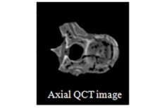
First of all, radiologic methods are used - in particular, review radiography, magnetic resonance [11] or computed tomography. Radiography helps to examine osteophytes of the lumbar spine, detect bony thickening and changes in the distance between the vertebrae. Computed tomography of the lumbar spine allows a detailed examination of the spine, detect stenosis of the spinal canal. Magnetic resonance imaging is more informative: thanks to this method, it is possible to visualize soft tissue structures (nerves, ligaments, discs), to detect compression of these structures.
If the doctor suspects damage to nerve fibers and endings, it will be appropriate to perform electroneuromyography - a method that allows you to find out the degree of nerve damage and disruption of nerve impulse conduction. In some cases, radioisotope scanning is additionally prescribed - a procedure based on the difference in the degree of absorption of radioactive materials by different tissues.
Differential diagnosis
Differential diagnosis is carried out with such pathologies:
- Kidney and urinary system diseases;
- Disorders of the gastrointestinal tract;
- Inflammatory processes in the pelvic organs;
- Injuries to the spine, internal organs, musculoskeletal system;
- Pelvic neurosis.
Among diseases of the kidneys and urinary tract should pay attention to the possible recurrence of chronic pyelonephritis or glomerulonephritis, urolithiasis. In the course of examination, in addition to general clinical and biochemical blood and urine tests, ultrasound, radiography (including contrast), computer or magnetic resonance imaging, puncture biopsy should be performed.
Among gastrointestinal pathologies should be excluded pancreatitis and cholecystitis, gallstones, irritable bowel syndrome, acute or chronic enterocolitis, Crohn's and Hirschprung's diseases, nonspecific ulcerative colitis, intestinal polyposis, tumors. In the process of diagnostics it is advisable to perform rectomanoscopy, fibrogastroduodenoscopy.
In addition, the possibility of neurinoma and other tumors involving the roots of spinal nerves should be excluded.
Who to contact?
Treatment of the osteophytes of the lumbar spine
When osteophytes of the lumbar spine appear, it is necessary to take measures to improve blood circulation, optimize the function of spinal muscles and metabolic processes. If there is an acute pain syndrome, the patient is prescribed bed rest. In general, treatment should include taking medication, massage, therapeutic exercise. LFK and manual therapy are relevant only during remission, when there is no acute pain.
Osteophytes are generally considered a degenerative condition and can be surgically removed by traditional or minimally invasive methods during spine surgery if they cause disability or neurological symptoms. [12]
In order to get rid of pain and other accompanying symptoms, to restore sensation and motor function, the following groups of drugs are prescribed:
- Painkillers (Ketorol, Ketanov, Metamizol);
- Non-steroidal anti-inflammatory drugs (Diclofenac, Ibuprofen, Depiofen).
In long-lasting pain, muscle spasm occurs. Myorelaxants (Midocalm, Sirdalud, Baklosan, Tolperisone) are used to relieve spastic muscle contraction.
In addition, drug therapy may include taking chondroprotectors (preparations with chondroitin and glucosamine), as well as vitamin and mineral complexes.
Surgical treatment is used if conservative methods are ineffective or if the patient develops severe neurological complications, for example:
- For limb paresis;
- When large intervertebral hernias form;
- When the spinal canal is severely narrowed;
- When the functionality of internal organs is impaired due to the progression of lumbar osteophytes;
- With intense pressure on the nerve endings;
- In case of massive osteophytes directly affecting the condition of nearby tissues.
The surgeon may use normalizing or compensatory interventions, such as laminectomy, facetectomy, foraminotomy, and so on.
A facetectomy is the removal of intervertebral articulations where osteophytes are found. If radicular syndrome is present, facetectomy is mandatory, sometimes combined with laminectomy. Facet articulations are removed under general anesthesia, by microsurgery and microscopy under radiographic control.
Foraminotomy is performed if there is a need to increase the intervertebral space to reduce compression of nerve roots. The surgery is performed in case of severe pain that cannot be corrected with medication, as well as in case of prolonged compression of the nerve outgrowth, impaired function of internal organs. During the intervention, the interfering bone part (osteophyte) is removed.
Microdiscectomy is the removal of the affected disc by microsurgery. The disc is not removed completely, but is preserved as much as possible by cutting away only the necessary tissue.
By performing laminectomy eliminate compression of nerve roots and spinal cord, removing spinous processes, intervertebral discs, vertebral outgrowths. Announcements of surgical interventions depend on the specific situation. [13]
Another method of mandatory use in osteophytes of the lumbar spine is physiotherapy. Physical procedures help to cope with pain syndrome, improve motor capabilities, eliminate muscle spasm, optimize blood circulation and lymph flow, stabilize the transmission of impulse signals along the nerves. In most cases, patients with lumbar osteophytes are recommended these procedures:
- Electromyostimulation;
- Magnetotherapy;
- Electro-pulse therapy;
- Electrophoresis with medications (corticosteroids, sulfur preparations, etc.);
- Laser therapy;
- Ultraviolet irradiation;
- UHF.
Physical therapy exercises are selected by a doctor, taking into account the course of the pathology, the patient's general state of health, body weight and physical fitness. Properly selected exercises help to strengthen muscles and ligaments, increase flexibility and mobility of the lumbar spine, improve blood circulation and metabolic processes, reduce pressure on intervertebral discs and vertebrae.
Prevention
To minimize the risks of osteophyte formation in the lumbar spine as much as possible, the following expert recommendations should be heeded:
- Keep physically active, do daily exercises, walk, swim;
- Make sure your posture is correct;
- If your work is predominantly sedentary, it is important to take regular breaks, get up, walk around, and warm up;
- To control your body weight;
- Eat a good diet, avoid overeating;
- Avoid injuries to the back and limbs, timely consult doctors about any disorders of the musculoskeletal system;
- Avoid overloading the spine (if you need to lift or carry a heavy object, you should do it correctly, with an even distribution of the load on the spinal column);
- Never start sports training without first warming up and warming up;
- Prevent the development of congestion and metabolic disorders;
- Use a quality comfortable bed (mattress, pillow) for night rest;
- See your doctor regularly for preventive checkups.
It is equally important to drink enough water and eat right. Specialists recommend completely avoiding or minimizing the amount of fast carbohydrates, alcoholic beverages, excessively salty, fatty and spicy dishes.
If possible, it is recommended to lead an active lifestyle, regularly perform morning exercises and visit the swimming pool, wear comfortable clothes and shoes.
Forecast
Prognosis in osteophytes of the lumbar spine depends on the degree of the pathological process, timeliness and quality of treatment. The disease is one of the common causes of disability, and in neglected situations, the patient may lose the ability to move and serve himself.
Patients with severe forms of osteophytes can receive the third or second form of disability, which depends on the stage and volume of pathology.
In general, provided competent and timely treatment, the prognosis can be considered favorable: osteophytes of the lumbar spine slow their growth, the patient's condition improves. Most often this can be achieved through conservative therapy with the use of nonsteroidal anti-inflammatory drugs and analgesics. Important: in order not to neglect the disease, it is necessary to contact doctors even at the appearance of the first signs of osteophytes. In complex cases, surgical intervention will be required to improve the condition.