Medical expert of the article

New publications
Marginal osteophytes of the hip joint
Last reviewed: 29.06.2025

All iLive content is medically reviewed or fact checked to ensure as much factual accuracy as possible.
We have strict sourcing guidelines and only link to reputable media sites, academic research institutions and, whenever possible, medically peer reviewed studies. Note that the numbers in parentheses ([1], [2], etc.) are clickable links to these studies.
If you feel that any of our content is inaccurate, out-of-date, or otherwise questionable, please select it and press Ctrl + Enter.
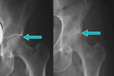
Often during radiography of the pelvic bones, marginal osteophytes of the hip joint are detected. These are specific pathological growths on the articular surface, where the bone is covered with cartilage. When in contact with nerve endings, osteophytes cause severe pain, mainly because of which patients seek medical help. The main reason for the appearance of growths is osteoarthritis and cartilage destruction. [1]
Epidemiology
Marginal osteophytes of the hip joint are more often found in men over 65 years of age. The age of 80% of patients - the overwhelming majority - exceeds 75 years.
For example, in the United States of America, the prevalence of the pathology is 12%, as a result of which several hundred thousand endoprosthetic surgeries are performed each year.
The most common manifestations of marginal osteophytes of the hip joint:
- Pain when trying to move and its absence at rest (sometimes irradiation to the groin area is noted);
- Transient stiffness in the joint in the morning;
- Limited range of motion in the hip joint, crepitation;
- Absence of signs of inflammation (swelling, local temperature rise).
Causes of the osteophytes of the hip joint.
In order to understand the causes of the formation of marginal osteophytes of the hip joint, it is necessary to have an idea about the anatomical and physiological features of this articulation.
The head of the femur and the acetabulum of the ilium are involved in the formation of the hip joint. The articular surfaces are covered with synovial cartilage tissue. This tissue can absorb synovial fluid and release it back when needed, depending on movement activity. When standing for long periods of time, the acetabulum is subjected to intense mechanical stress. During walking, the cushioning load shifts depending on the change in the motor vector. In this situation, different and changing load directions affect almost the entire surface of the acetabulum and femoral head.
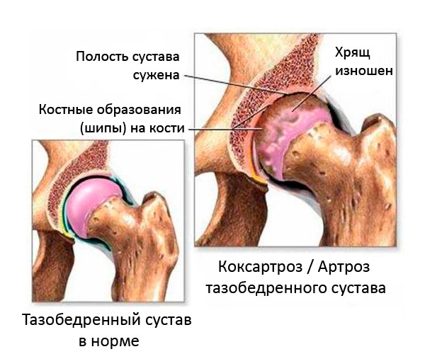
The formation of marginal osteophytes occurs only when the synovial layer of cartilage is damaged. In areas of cartilage thinning and bone exposure during motor activity, microcracks form in the cartilage, which over time become filled with calcium salts. Such deposits damage the soft tissues, which leads to a constant inflammatory process. As a result, cartilage tissue is destroyed, osteophytes grow and further throughout the entire inner articular surface. [2]
Indirect causes of this phenomenon can be:
- Overweight, which repeatedly increases the load on the surfaces of the hip joint and provokes accelerated destruction of the cartilage layer;
- Sedentary lifestyle;
- Curvature of the lower limbs and feet;
- Injuries to the hips and the hip joint itself;
- Spinal curvature with pelvic misalignment, knee arthrosis with misalignment and limb shortening;
- Age-related biochemical changes;
- Growth anomalies (juvenile epiphyseolysis of the femoral head, infantile osteonecrosis);
- Skeletal anomalies (dysplasia of the hip joint or acetabulum, rotational malalignment of the femoral neck);
- Femoral-acetabular impingement (elevation in the anterior external segment of the head-neck junction, excessive sheltering of the femoral head by the acetabulum);
- Epiphyseal anomalies (spondyloepiphyseal dysplasia);
- Hormonal disorders (low estrogen levels in women).
Risk factors
Since the pathogenesis of the appearance of marginal osteophytes of the hip joint is not fully understood, it is important to know about the risk factors that can provoke the development of pathology. Such factors include:
- Obesity, overweight, increasing the load on the joint surfaces and causing accelerated destruction of cartilage tissue;
- Sedentary lifestyle (predominantly sedentary work, hypodynamia due to excess weight, etc.);
- Foot malposition, bone deformities (including valgus curvature);
- Traumatic injuries to the hip joint or upper thigh;
- Sacro-lumbar osteochondrosis;
- Incorrect posture, deformations of the spinal column, which entails uneven distribution of the shock-absorbing load during motor activity;
- Regular heavy physical activity with prolonged stay "on your feet", manual transportation of heavy objects;
- Diseases of the vessels of the lower extremities (varicose veins, angiopathy of diabetic origin, obliterating endarteritis, atherosclerosis, etc.);
- Rheumatoid damage to intra-articular cartilage due to rheumatoid arthritis, gout, Bechterew's disease (joint type), systemic lupus erythematosus, etc.;
- Improper lifestyle, poor diet, low fluid intake during the day.
In the elderly, marginal osteophytes can occur as a result of trauma, fractures in the area of the femoral head. In middle-aged patients, it is necessary to exclude all kinds of endocrine disorders that can cause cartilage destruction.
High-risk groups include women during pregnancy (late pregnancy is characterized by physiological softening of cartilage tissue), as well as obese people.
Pathogenesis
The pathogenetic picture of the formation of marginal osteophytes of the hip joint is still being studied. It is known that in most cases osteophytes occur at a late stage of osteoarthritis development: the growths are localized on the femoral head or on the surface of the acetabulum of the iliac bone.
Topographical, morphological and other features of osteophyte growth were first described in 1975. At the same time, the classification of growths depending on their location and growth was determined. In particular, marginal osteophytes were divided into peripheral (with localization along the edge of the femoral head) and centralized (with localization along the edge of the rough fossa of the femoral head). In addition to marginal osteophytes, episarticular and subarticular osteophytes have also been described.
Variants of osteophyte growth:
- There is an overgrowth of broad and flat osteophytes affecting the medial and posterior zones of the femoral head, with preservation of sphericity. Sometimes there are degenerative changes with cystic formations in the anterior superior and medial segment of the femoral head. Clinical and radiologic examination reveals lateral rotation and displacement of the femoral head in relation to the acetabulum.
- The growths tend to spread outward and affect the posterior and medial areas of the femoral head. The bone tissue is destroyed, the upper and lateral areas of the femoral head are involved, and the femoral head is displaced laterally and upwards relative to the acetabulum. Clinical signs are fixed flexion contracture, lateral rotation, and hip adduction.
- The marginal osteophytes of the surfaces of the acetabulum and femoral head form a peculiar ring surrounding the hip articulation. There are destructive and degenerative changes in the medial and posteromedial region of the femoral head.
- Peripheral marginal osteophytes become visible when the acetabulum with the femoral head is deeply recessed to the pelvic side. As bone destruction progresses, the head is displaced upward relative to the acetabulum, and a ring of peripheral growths is found along the inferior edge of the femoral head.
Symptoms of the osteophytes of the hip joint.
Symptoms of the formation of marginal osteophytes of the hip joint may not manifest themselves immediately after the onset of pathological changes. Only over time, as they grow, there is constant pain in the hip joint and limitation of movement.
Many patients suffering from marginal osteophytes of the hip joint complain of pain in the lower back, buttocks and hips. The pain syndrome can range from minor discomfort to acute severe pain. In advanced cases, the pain is so severe that the patient is unable to make any movements.
Freedom of movement in the joint is also impaired. Constant discomfort and pain make it difficult to perform even simple movements: it becomes problematic to walk, lift the leg or even sit for long periods of time. Many people have a feeling of stiffness in the joint, a feeling that "the leg does not obey".
Edge osteophytes of the hip joint is a frequent pathology that cannot be cured completely. However, timely referral to doctors when the first symptoms are detected helps to start treatment in time and prevent the development of serious consequences. [3]
Complications and consequences
Degenerative-dystrophic joint pathologies with subsequent formation of osteophytes is not only a medical, but also a social problem, as patients in many cases become disabled. The main consequence of the formation of marginal osteophytes of the hip joint is the loss of the ability to lead a normal lifestyle. At first, the patient experiences discomfort when walking for a long time. After a while, it becomes necessary to make stops while walking (almost every 200-300 m), then it becomes necessary to use a support cane or crutches.
Due to tissue destruction and overgrowth of marginal osteophytes, the patient experiences severe pain, the ability to perform movements is severely limited. Pathological processes contribute to the development of chronic inflammation in the joint and surrounding tissues, arthritis or periarthritis, osteomyelitis occur.
The muscles of the affected lower limb atrophy, the leg becomes noticeably thinner. The imbalance of load leads to disorders of other components of the musculoskeletal system: flat feet, osteochondrosis, deformed spinal column, nervous system suffers (compression neuropathies, etc.).
Among the no less serious consequences are the formation of pathological subluxations, ankylosis (fusion of the joint surfaces), and necrosis. As a result, the patient becomes disabled and loses the ability to move independently. The risks of congestion, thrombosis, etc. Increase.
In advanced cases, the only possible way to improve the situation is endoprosthesis - a complex surgical intervention, associated with a high risk of complications and a large number of contraindications. Therefore, it is important to seek medical attention in a timely manner: early treatment can slow down or stop the progression of painful processes without resorting to major surgery.
Diagnostics of the osteophytes of the hip joint.
During the initial consultation, the doctor collects anamnesis, externally assesses the state of the musculoskeletal system, examines and feels the affected joints. To clarify the nature of pathological changes in the central nervous system, a general neurological examination is performed.
Comprehensive instrumental diagnostics may include:
- Radiography of the hip joints in several projections, with determination of the type and location of osteophytes;
- Computer or magnetic resonance imaging to determine the stage of the disease, clarify the features of the growths, detail and study all the structures involved;
- Ultrasound of soft tissues, joints;
- Electroneuromyography to assess the functionality of the nervous system in peripheral regions.
If necessary, the doctor may resort to additional diagnostics to obtain more precise information about the condition of the hip joint and the marginal osteophytes. In particular, arthroscopy or biopsy is used.
Additionally, laboratory tests are prescribed:
- Hemogram is performed to detect markers of inflammation (increased COE and leukocytes);
- Blood biochemistry is performed to find out the level of calcium, C-reactive protein, rheumatoid factor;
- Serologic screening is necessary to determine specific immunoglobulins and autoimmune antibodies.
If the patient has systemic diseases or other indications, then consultations with an endocrinologist, traumatologist, rheumatologist, etc. Are prescribed.
Differential diagnosis
Differential diagnosis is performed with the following pathologies:
- Osteonecrosis.
- From the initial stage of osteonecrosis to the late stages, the femoral head gradually flattens, with no pathologic changes in the joint itself.
- Osteoarthritis is only detected at a late stage of osteonecrosis.
- Femoral acetabular impingement.
- Femoral impingement syndrome of the anterior external segment of the cephalic-neck junction (cam impingement).
- Impingement of the anterosuperior segment of the acetabulum (pincer impingement).
- Hip dysplasia.
- External flattening of the acetabulum.
- Pyrophosphate arthropathy.
- Pyrophosphate deposits in the acetabular lip and cartilage.
- Degenerative changes in the hip joint, formation of osteophytes.
- Subchondral cysts.
Treatment of the osteophytes of the hip joint.
Medication for marginal osteophytes of the hip joints includes the use of analgesics and anti-inflammatory drugs. Analgesics (Ketonal, Dexalgin, Nalgesin) will help to reduce pain and improve the quality of life of the patient, and anti-inflammatory drugs (Diclofenac, Paracetamol, Ibuprofen) will stop the development of inflammatory reaction.
Special chondroprotective drugs help slow the progression of osteoarthritis, which often precedes the formation of marginal osteophytes. Chondroprotectors promote regeneration of cartilage tissue, improve joint mobility. However, such drugs (Glucosamine, Chondroitin sulfate) require long-term use, as they have an accumulative effect.
Myorelaxants are another group of medications useful for patients with marginal osteophytes of the hip joint. These medications reduce muscle tension, improve mobility, and relieve pain. Among the most common myorelaxants are: Midocalm, Tizanidine, Baclofen.
In general, a comprehensive treatment consisting of conservative and surgical methods is used.
Physiotherapeutic treatment includes the use of electrophoresis and ultraphonophoresis, allowing to eliminate muscle spasms, relieve pain, improve metabolic processes in tissues.
Physical therapy is another important therapeutic component. Physical therapy exercises are also prescribed during rehabilitation, which is necessary to strengthen the muscles of the hip region and lower limbs.
Acupuncture and manual therapy sessions are also recommended to relieve muscle tension and pain.
The use of special orthopedic constructions (inserts, insoles, orthoses) is indicated in case of deformities, different limb lengths, etc.
Modern surgical technologies often help to slow down the progression of marginal osteophyte formation and eliminate the need for endoprosthetics. Thus, endoscopic interventions are performed on the hip joint - arthroscopy with replacement of damaged tissues. The operation is performed through small skin incisions (punctures). Optics and endoscopic instruments are inserted into the joint, and a special monitor provides an opportunity to examine in detail all pathologically altered joint tissues. With the help of instruments and under the control of optics, osteophytes of the femur and acetabulum are removed, and the articular lip is sutured. If the articulation is deformed, it is given an anatomically correct configuration. Damaged cartilage is replaced with a collagen biomatrix, which is fully capable of performing the function of normal cartilage tissue.
As for endoprosthetics, this intervention is appropriate when the hip joint is completely and irreversibly dysfunctional and cannot be repaired. During endoprosthetic surgery, the surgeon replaces the affected joint surface with an artificial one.
After surgical intervention, the patient undergoes prolonged rehabilitation with physiotherapy and physical therapy. The recovery period can take several months and requires not only patience, but also considerable effort on the part of the patient, including strict adherence to all medical recommendations.
Prevention
Preventive measures should include preventing the development of osteoarthritis and maximizing cartilage preservation.
The diet should contain collagen, which is necessary to support joint function and structure. Collagen is present:
- In meat and fish broth;
- In cold cuts, jelly;
- In berries, fruits, vegetables.
It is necessary to consult with rehabilitation physicians or physical therapy instructors about physical therapy. For each specific case, a different set of exercises is selected.
Recommended:
- Regular massage courses (1-2 times a year);
- Treatment and prevention of metabolic diseases (obesity, diabetes, gout), as well as pathologies of the digestive tract and liver;
- Correction of foot curvature, use of orthopedic shoes and special insoles;
- Providing the body with the necessary vitamins and trace elements, additional intake of vitamin D, magnesium, zinc;
- Prophylactic administration of chondroprotectants;
- Avoiding injuries and excessive loads on the lower limbs and hip joints in particular;
- Observance of the labor and rest regime;
- Regular medical examinations for the timely detection of pathologies of the musculoskeletal system.
Forecast
The initial stage of the formation of marginal osteophytes usually does not lead to disability. It is important to see a doctor in time, have a full examination, start treatment, and follow all medical recommendations.
The prognosis is considered less favorable when it comes to neglected cases, a large overgrowth of osteophytes, especially in secondary osteoarthritis. The disease is prone to rapid progression, the hip joint is quickly destroyed. For several years, the patient may become disabled.
In complicated cases, it may be necessary to undergo complex endoprosthetic surgery. Modern treatment methods help people to return to their normal way of life.
Unfortunately, in most cases, patients do not immediately seek medical help, so the disease quickly progresses, the joints are deformed. Over time, marginal osteophytes of the hip joint lead to severe pain and disability.
Literature used
Application of injectable forms of biopolymer heterogeneous hydrogels in degenerative-dystrophic lesions of joints, Practical Manual for Doctors, Moscow, 2012
Modern approach to pathogenesis, diagnosis and treatment of osteoarthritis of the knee joint E.M. Lisitsyna, M.P. Lisitsyn, A.M. Zaremuk
Traumatology and Orthopedics, Ryabchikov I.V. Kazan, 2016