Medical expert of the article

New publications
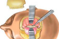
Craniotomy
Last reviewed: 29.06.2025

All iLive content is medically reviewed or fact checked to ensure as much factual accuracy as possible.
We have strict sourcing guidelines and only link to reputable media sites, academic research institutions and, whenever possible, medically peer reviewed studies. Note that the numbers in parentheses ([1], [2], etc.) are clickable links to these studies.
If you feel that any of our content is inaccurate, out-of-date, or otherwise questionable, please select it and press Ctrl + Enter.
Craniotomy is a neurosurgical intervention used in medicine since ancient times. Today, the operation involves the use of microsurgical instruments, a special microscope, power devices, so the technological capabilities of craniotomy have increased significantly. Obtaining comprehensive information about the anatomy, pathogenesis of various lesions, about the possibilities of using this or that instrumentation, about the technique and the main stages of opening the cranium has led to a significant reduction in the risks of complications. [1]
The term "craniotomy" literally means "cranial incision" in Greek. It is a neurosurgical operation in which the surgeon makes a hole in a certain place in the skull bone to provide access to the brain, brain membranes, vessels, tumors, etc. In addition, this procedure helps to reduce the progression of intracranial pressure, thereby preventing the formation of complications, structural displacement of the brain, and related fatalities. [2]
Indications for the procedure
Craniotomy surgery involves removing a segment of cranial bone to provide access to the brain with further bone replacement. The intervention is often used in neurosurgery for aneurysms and tumor intracerebral processes.
The operation is indicated for benign or malignant brain tumors. In the case of malignant tumors, biopsies can be taken and part or all of the tumor can be resected during the craniotomy.
Intervention is performed in cases of cerebral vascular diseases (aneurysms or arteriovenous malformations), craniocerebral trauma (fractures and hematomas), intracerebral infection (abscesses, etc.), neurological pathologies, including severe epilepsy.
Craniotomy is indicated for primary neoplasms: [3]
- Benign (Meningioma );
- Malignant (Glioma ). [4]
Surgery is possible for germinomas and lymphomas, brain metastases.
In general, specialists distinguish such indications for intervention:
- Removal of a benign or malignant mass that puts pressure on the brain, which leads to headaches, disorders of consciousness, disturbances in orientation in space;
- Repairing vascular defects; [5]
- Repairing a skull fracture, brain hemorrhage;
- Treatment of an intracerebral infectious process;
- Treatment of neurological pathologies, severe epilepsy;
- Correction of anomalies or distortions of the cranium in children.
Craniotomy in most cases helps to alleviate the symptoms of the pathology. However, it is important to realize that the intervention involves opening the skull and exposing the brain, which is a technically complex neurosurgical manipulation.
Preparation
Like any other surgery, craniotomy requires multiple levels of diagnosis beforehand. [6] Patients are prescribed:
- Electrocardiography or other cardiac diagnostics (depending on the indications and age of the patient);
- X-rays of the lungs (fluoroscopy or review);
- A CT scan of the skull;
- Magnetic resonance imaging or functional MRI;
- Cerebral angiography with contrast;
- Positron emission tomography or positron emission computed tomography (in case of metastasis); [7]
- CT angiography.
The surgeon carefully studies the patient's medical history, previous diseases, the presence of hereditary predisposition. It is obligatory to keep a record of drugs used in treatment, which allows the anesthesiologist to correctly determine the nature and dose of anesthesia. [8], [9]
Approximately 8 hours before the intervention, you should not eat or even drink any liquids, including water. It is advisable to refrain from smoking.
Immediately before surgery, jewelry, dentures, lenses, etc. Should be removed.
If the patient has taken any medications, it is necessary to tell the doctor about it. Drugs affecting blood clotting processes are discontinued no later than 7 days before the expected date of craniotomy.
Any additional examinations can be ordered on a case-by-case basis to clarify individual points when planning a craniotomy. [10]
Craniotomy instruments
Specialized equipment is required to perform a craniotomy.
The operating table should ensure a stable position of the operated patient. There must be an automated mechanism capable of transforming the position of the table and its individual parts depending on the operational requirements, for the convenience of a particular access.
The patient's head must be rigidly secured - e.g. With a Mayfield 3-point brace. The neurosurgical instruments must be comfortable, suitable for use in confined spaces, and at the same time functionally simple.
In most cases, tool kits like this are used:
- Common neurosurgical instruments:
- Blunt-ended straight bipolar;
- Aspirators;
- A set of clips with an overlay;
- Novocaine or lidocaine with adrenaline in a syringe;
- A peritoneal scalpel;
- Tweezers;
- Jantzen's wound dilator;
- Scissors;
- Retractor.
- Craniotomy instruments:
- Rotary cutters; [11]
- Raspator;
- Folkman's spoon;
- Polenov's guide with a Jiggly Olivecrown saw;
- Bone cutters and Kerrison's;
- Scalpel;
- Scissors to dissect the dura mater.
A perforator, craniotome with dura protection, speed handle and diamond burr may also be required.
Technique of the craniotomies
Before craniotomy, the patient's hair is shaved off in the area of the proposed intervention. The skin is treated with antiseptic solution.
Fixation of the patient's head on the operating table is an important moment for the success of the intervention. The head should be elevated and rotated relative to the trunk, avoiding excessive bending of the neck and associated impairment of venous circulation and increased intracranial pressure.
Subsequent stages of craniotomy involve preparation of the surgical field within the known rules of asepsis and antisepsis. General anesthesia is used for the vast majority of such operations.
The execution of the incision line depends on the location and configuration of the bone flap and the features of the vascular and nervous network in the operated area. The base of the flap is directed to the cranial base, to the main feeding vessels, which helps prevent ischemia and necrosis of soft tissues.
Before making the incision, the neurosurgeon may perform soft tissue hydropreparation to stop bleeding and improve the mobility of the cutaneous aponeurotic segment. Novocaine infiltration along the incision border has been successfully used for this purpose. If there are no contraindications, the use of adrenaline may be recommended to spasm arterial vessels and prolong the effect of novocaine.
Bleeding is stopped using special skin clips with capture of both the vessel and the skin-aponeurotic segment. The emissary vessels are blocked with wax or Luer's cutters by pinching the external and internal bone plates, pinching the trabeculae.
The bony periosteal fragment is isolated by cutting the periosteum in an arc-shaped manner using a scalpel, with an indentation of 10 mm from the border to the center. The periosteum is peeled away from the incision to a distance corresponding to the diameter of the cutter.
The classic variant of craniotomy today is the formation of a free bone flap with a craniotome on the basis of a single cutter hole. The dura mater is opened by making a cruciform or horseshoe-shaped incision. Vessels are coagulated before opening, as the dura is much more difficult to suture in a wrinkled curved form. Further intervention is carried out depending on its intended focus. [12]
At the end of the operation, the wound is closed in layers using a three-row suture. Depending on the situation, subdural, epidural, or subgaleal passive drainage is used. Sutures are removed on 8-10 days.
The average duration of a craniotomy is 2.5-3 hours, depending on the extent and complexity of the surgery. Sometimes more than 4 hours may be required.
Several types of craniotomies are known:
- Decompressive craniotomy (combined with removal of hemorrhage inside the skull to stabilize and control intracranial pressure - e.g. In craniocerebral trauma). [13], [14]
- Resection craniotomy (involves partial resection of bone tissue).
- Bone-plasty craniotomy (involves placement of a previously removed bone, dural-bone-periosteal, or skin-muscle-periosteal-bone flap in its original place).
- Stereotactic craniotomy (performed under the control of magnetic resonance or computerized tomography).
- Endoscopic craniotomy (accompanied by insertion of an endoscopic device with light and camera through the bone opening).
- "Keyhole" (a low-damage procedure that involves making a small hole in the behind-the-ear area - primarily used to remove neoplasms).
- Craniotomy "awake" (sedation and local anesthesia are used instead of general anesthesia). [15]
- Suboccipital craniotomy (performed in the area of the large (cerebellopontine) cistern of the brain).
- Supraorbital (the so-called "brow craniotomy" is used to remove forebrain neoplasms).
- Pterional, or frontal temporal craniotomy (involves making an incision in the temporal region along the line of hair growth - specifically in the wing-shaped cranial zone). [16], [17], [18]
- Orbitozygomatic craniotomy (suitable for removal of aneurysms and complex neoplasms, performed along the curve of the orbital line).
- Posterior fossa craniotomy (involves making an incision at the base of the skull).
- Translabyrinthine craniotomy (accompanied by partial removal of the mastoid process and semicircular canals).
- Bifrontal craniotomy (used to resect solid neoplasms in the front of the brain).
Depending on the focus of intervention and the peculiarities of the pathology, the neurosurgeon selects the surgical access that is optimal for a particular case. In particular, a Kozyrev craniotomy may be used. During the operation, a part of the cranial bone (the so-called bone flap) is separated from the rest of the skull to gain access to the structures closed to visualization (dura mater, brain, nerves, vessels, etc.). Craniotomy and craniectomy involve the use of special instruments described above. After the intervention, the surgeon replaces the bone flap with appropriate titanium plates, attaching them to the surrounding part of the bone with screws. If the bone segment is removed but not replaced immediately, this procedure is called trepanation. It is performed when there is an increased risk of cerebral edema or when a one-stage bone flap replacement is not possible.
Thus, the only difference in the terms craniotomy and trepanation is whether the formed bone defect is replaced immediately or after a period of time. In both cases, the surgeon makes a hole in the skull bone to gain access directly to the brain tissue.
Interventions can vary in size and complexity. Small craniotomies of approximately 19 mm are referred to as "burrs" and openings of 25 mm or more are called "keyholes". These types of accesses are used for minimally invasive procedures such as:
- To shunt the cerebral ventricle to drain the liquor in hydrocephalus;
- For deep brain stimulator placement, endoscopy;
- To monitor intracranial pressure readings; [19]
- For puncture biopsy, hematoma aspiration.
Complex craniotomy is performed on patients with severe pathologies:
- With brain tumors;
- Subdural or epidural hematomas, hemorrhages;
- Abscesses;
- With vascular aneurysms;
- Epilepsy, dura damage. [20]
Craniotomy is also used for microvascular decompression of the trigeminal nerve ending in patients with neuralgia.
Fetal craniotomy
Separate mention should be made of the so-called fetal-destroying operations - obstetric interventions involving the destruction of the fetus with its further removal through the birth canal. Such manipulations are carried out if there is a threat to the life of the mother, mainly when the fetus has already died, to ensure the possibility of its extraction and saving the life of the woman against the impossibility of using for any reason other techniques of obstetrics.
In this case, craniotomy involves the destruction and removal of the fetal brain through a hole made in its cranial box, allowing the head to be reduced in size by excerebration or cranioclasia.
For such an intervention, the kephalotribe is used - a surgical instrumentation, which is a strong forceps with which the doctor grasps the perforated head in order to subsequently remove the fetus in the course of fetus-destroying surgery.
Indications for this intervention may include:
- Fetal hydrocephalus;
- Frontal, antero-facial presentation;
- Threat of uterine rupture;
- Pinching of the soft tissues of the birth canal;
- Severe condition of a woman in labor, acute need for immediate delivery.
In the vast majority of cases, the operation is performed in the case of fetal death, or defects and pathologies that make further existence of the child impossible.
Contraindications to the procedure
Age and most chronic diseases most often do not become contraindications to craniotomy. Skilled surgeons operate on patients of almost any age.
Surgery may be contraindicated in the acute period of infectious-inflammatory processes, in general severe decompensated state. In such cases, the possibility of performing manipulation is determined individually, separately for each specific situation.
Craniotomy may be indicated after appropriate therapy has been administered.
Complications after the procedure
Before a craniotomy is scheduled, the patient and their loved ones are told about the possible complications of this complex neurosurgical operation.
To minimize the risks, it is important to provide the operating doctor and anesthesiologist with all anamnestic information in advance. Only on the basis of mutual trust can all aspects of the upcoming intervention be optimally defined and adjusted.
Surgical complications of craniotomy are considered to be: [21]
- Wound infection;
- Bleeding;
- Cerebral edema;
- Disruption of the integrity of nearby vessels and tissues;
- Seizures.
According to statistical data, severe consequences after the procedure are relatively rare - no more than 4% of cases. These include partial or complete paralysis, amnesia, loss of speech or cognitive abilities. Fatal outcomes are reported in no more than 2% of cases.
To minimize risks, many patients receive certain treatments before or after surgery - for example, to reduce fluid buildup in brain tissue. Possible side effects include:
- Drowsiness or insomnia;
- Change in appetite;
- Muscle weakness;
- Weight gain;
- Digestive disorders;
- Irritability, mood swings.
If a seizure syndrome occurs, the patient may be treated with anticonvulsants.
Immediately after the craniotomy, swelling and bruising may occur in the face and near the eyes. In most cases, these effects disappear on their own within a few days.
Pain for a few days after the intervention cannot be ruled out, [22] which can be relieved by taking analgesics. Nausea is also possible, sometimes to the point of vomiting.
The most common consequences of craniotomy: [23]
- Visible scars;
- Facial nerve damage;
- Seizures;
- Weakness in certain muscle groups;
- Formation of a small depression in the area of intervention;
- Damage to the paranasal sinuses;
- Speech impediments, memory problems;
- Vestibular Disorders;
- Blood pressure instability;
- The body's reaction to anesthesia.
Relatively rare complications include strokes, blood clot formation, pneumonia, coma and paralysis, attachment of infectious processes, and cerebral edema. [24], [25]
Care after the procedure
Craniotomy is a serious surgical intervention in the brain area and therefore requires complex and lengthy rehabilitation measures. The primary rehabilitation period lasts several days and depends on the type of anesthesia used. At the postoperative stage, the patient must remain in the medical institution under the constant supervision of medical specialists. If there is instability or complications, the patient may be kept in the intensive care unit for several days.
The patient is discharged after about 1-1.5 weeks, depending on individual performance and the speed of recovery of the body.
For two months after craniotomy, driving vehicles and working with complex mechanisms should be avoided. Return to normal life activity is possible only after the disappearance of dizziness and pain in the head, recovery of functional abilities of the body.
It is imperative to see a doctor if:
- Vestibular, coordination and muscle strength disorders;
- Mental state has changed (memory and thinking processes have deteriorated, reactions have weakened);
- Pain, redness, bleeding or other discharge from the surgical incision area;
- I have a constant headache;
- Developed torticollis (a disorder of the musculoskeletal apparatus of the neck);
- Vision is impaired (blurred vision, "flies", double images, etc.);
- Seizures, impaired consciousness;
- Numbness, tingling, sharp weakness in the face, extremities;
- Symptoms of an infectious disease (fever, chills, brokenness, etc.);
- Nausea and vomiting that does not disappear after taking the prescribed medication for 2 or more days;
- There is pain that is not relieved by taking prescribed analgesics;
- Chest pains, shortness of breath, coughing;
- Problems with urinary control, stool control;
- Signs of lower extremity thrombosis (swelling, pain, fever, hyperemia of the legs).
Testimonials
In the vast majority of cases, craniotomy provides a permanent improvement in the patient's condition, depending on the pathology and the reason for the operation. The surgical technique is complex, but the results almost always meet expectations. If the procedure was performed for a neoplasm that caused severe and persistent headaches, they usually disappear after the operation.
In case of weakness or paralysis of the limbs, which is due to compression of the brain by the neoplasm, the patient's condition usually improves.
When the tumor process invades the brain tissue, the prognosis is less optimistic.
Craniotomy often helps to eliminate epileptic seizures, but it is important to know that in some cases this does not happen or the situation worsens.
Surgery alone or in conjunction with radiation can control or cure many types of neoplasms, including astrocytomas, ependymomas, gangliogliomas, meningiomas, and craniopharyngiomas. Invasive tumors - particularly anaplastic astrocytomas, glioblastomas - are often not curable. However, in many cases it is possible to first perform surgical reduction of the size of the neoplasm and further neutralize it by radiation and chemotherapy. If it is not possible to remove the entire tumor process, it is often possible to improve the patient's well-being and prolong his life.
Craniotomy allows successful removal of benign brain neoplasms without subsequent recurrence.
Sources
- González-Darder JM. [History of the craniotomy]. Neurocirugia (Astur). 2016 Sep-Oct;27(5):245-57.
- Subbarao BS, Fernández-de Thomas RJ, Eapen BC. StatPearls [Internet]. StatPearls Publishing; Treasure Island (FL): Aug 1, 2022. Post Craniotomy Headache.
- Bhaskar IP, Zaw NN, Zheng M, Lee GY. Bone flap storage following craniectomy: a survey of practices in major Australian neurosurgical centers. ANZ J Surg. 2011 Mar;81(3):137-41.
- Schizodimos T, Soulountsi V, Iasonidou C, Kapravelos N. An overview of management of intracranial hypertension in the intensive care unit. J Anesth. 2020 Oct;34(5):741-757.
- Sahuquillo J, Dennis JA. Decompressive craniectomy for the treatment of high intracranial pressure in closed traumatic brain injury. Cochrane Database Syst Rev. 2019 Dec 31;12(12):CD003983.
- Alkhaibary A, Alharbi A, Alnefaie N, Oqalaa Almubarak A, Aloraidi A, Khairy S. Cranioplasty: A Comprehensive Review of the History, Materials, Surgical Aspects, and Complications. World Neurosurg. 2020 Jul;139:445-452.
- Buchfelder M. From trephination to tailored resection: neurosurgery in Germany before World War II. Neurosurgery. 2005 Mar;56(3):605-13; discussion 605-13.
- Andrushko VA, Verano JW. Prehistoric trepanation in the Cuzco region of Peru: a view into an ancient Andean practice. Am J Phys Anthropol. 2008 Sep;137(1):4-13.
- Enchev Y. Neuronavigation: geneology, reality, and prospects. Neurosurg Focus. 2009 Sep;27(3):E11.
- Hobert L, Binello E. Trepanation in Ancient China. World Neurosurg. 2017 May;101:451-456.
- Rao D, Le RT, Fiester P, Patel J, Rahmathulla G. An Illustrative Review of Common Modern Craniotomies. J Clin Imaging Sci. 2020;10:81.
- Sperati G. Craniotomy through the ages. Acta Otorhinolaryngol Ital. 2007 Jun;27(3):151-6.
- Yasargil MG, Antic J, Laciga R, Jain KK, Hodosh RM, Smith RD. Microsurgical pterional approach to aneurysms of the basilar bifurcation. Surg Neurol. 1976 Aug;6(2):83-91.
- Yaşargil MG, Reichman MV, Kubik S. Preservation of the frontotemporal branch of the facial nerve using the interfascial temporalis flap for pterional craniotomy. Technical article. J Neurosurg. 1987 Sep;67(3):463-6.
- Hendricks BK, Cohen-Gadol AA. The Extended Pterional Craniotomy: A Contemporary and Balanced Approach. Oper Neurosurg (Hagerstown). 2020 Feb 01;18(2):225-231.
- Choque-Velasquez J, Hernesniemi J. One burr-hole craniotomy: Lateral supraorbital approach in Helsinki Neurosurgery. Surg Neurol Int. 2018;9:156.
- Choque-Velasquez J, Hernesniemi J. One burr-hole craniotomy: Subtemporal approach in helsinki neurosurgery. Surg Neurol Int. 2018;9:164.
- Zieliński G, Sajjad EA, Robak Ł, Koziarski A. Subtemporal Approach for Gross Total Resection of Retrochiasmatic Craniopharyngiomas: Our Experience on 30 Cases. World Neurosurg. 2018 Jan;109:e265-e273.
- Zhou C, Evins AI, Boschi A, Tang Y, Li S, Przepiorka L, Sadhwani S, Stieg PE, Xu T, Bernardo A. Preoperative identification of the initial burr hole site in retrosigmoid craniotomies: A teaching and technical note. Int J Med Robot. 2019 Jun;15(3):e1987.
- Stachniak JB, Layon AJ, Day AL, Gallagher TJ. Craniotomy for intracranial aneurysm and subarachnoid hemorrhage. Is course, cost, or outcome affected by age? Stroke. 1996 Feb;27(2):276-81.
- Legnani FG, Saladino A, Casali C, Vetrano IG, Varisco M, Mattei L, Prada F, Perin A, Mangraviti A, Solero CL, DiMeco F. Craniotomy vs craniectomy for posterior fossa tumors: a prospective study to evaluate complications after surgery. Craniotomy vs. Craniectomy for posterior fossa tumors: a prospective study to evaluate complications after surgery. Acta Neurochir (Wien). 2013 Dec;155(12):2281-6.
- Hamasaki T, Morioka M, Nakamura H, Yano S, Hirai T, Kuratsu J. A 3-dimensional computed tomographic procedure for planning retrosigmoid craniotomy. Neurosurgery. 2009 May;64(5 Suppl 2):241-5; discussion 245-6.
- Broggi G, Broggi M, Ferroli P, Franzini A. Surgical technique for trigeminal microvascular decompression. Acta Neurochir (Wien). 2012 Jun;154(6):1089-95.
- Alvis-Miranda H, Castellar-Leones SM, Moscote-Salazar LR. Decompressive Craniectomy and Traumatic Brain Injury: A Review. Bull Emerg Trauma. 2013 Apr;1(2):60-8.
- Dreval, Baskov, Antonov: Neurosurgery. Manual for physicians. In 2 volumes. Volume 1, Publisher: GEOTAR-Media, 2013.