Medical expert of the article
New publications
Calcinates in the breast
Last reviewed: 12.07.2025

All iLive content is medically reviewed or fact checked to ensure as much factual accuracy as possible.
We have strict sourcing guidelines and only link to reputable media sites, academic research institutions and, whenever possible, medically peer reviewed studies. Note that the numbers in parentheses ([1], [2], etc.) are clickable links to these studies.
If you feel that any of our content is inaccurate, out-of-date, or otherwise questionable, please select it and press Ctrl + Enter.

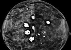
In recent years, calcifications in the mammary gland (deposition of calcium salts) have been diagnosed many times more often than before.
If breast disease is suspected, a woman is recommended to undergo a number of examinations, among which mammography is considered the most effective. Calcifications in the mammary gland can be determined using mammography, as well as other types of X-ray examinations.
Causes of calcifications in the breast
There may be several causes of calcifications in the mammary gland, the most dangerous being breast cancer. In any case, when microcalcifications are detected, the patient is immediately prescribed a biopsy. Causes of calcium salt deposits in the breast:
- breast cancer;
- overdose when taking calcium and vitamin D3;
- lactation stagnation;
- salt deposition;
- menopause;
- metabolic disorders;
- age-related changes in the body.
Pathogenesis
Mammologists note that, as a rule, calcium salt deposits do not pose a threat to a woman's health and life. They are formed due to metabolic disorders, stagnation during prolonged lactation, overdose of vitamin D3 and calcium, during menopause. However, about 20% of cases of this pathology are caused by breast cancer. Therefore, a thorough diagnosis is absolutely necessary when this pathology is detected; this process should not be left to chance under any circumstances. After calcium salt deposits are detected, a biopsy is immediately prescribed.
Microcalcifications can be found in the ducts of the breast, lobules and stroma of the breast. As a rule, lobular foci of pathology are benign, they occur with fibrocystic mastopathy, breast cysts, metabolic disorders, and do not require treatment.
Symptoms of calcifications in the breast
Microcalcifications are not palpable, which means that they cannot be determined during examination. Calcium salt deposits are determined during mammography, which should be done once a year. Symptoms of calcifications in the mammary gland are usually absent. It is for this reason that pathology at an early stage of development is quite difficult to detect; a woman should do preventive mammography at least once a year.
The presence of calcium salt deposits in the body can be shown by a biochemical blood test and a blood test for hormones.
 [ 12 ]
Where does it hurt?
Forms
Calcium salt deposits can have different shapes and sizes, be single or multiple. They are divided into lobular, ductal, stromal.
Single calcifications in the mammary gland
There are a number of reasons for the appearance of calcifications in the mammary gland. They are difficult to diagnose, so you need to do a mammogram regularly, once a year. Microcalcifications in the breast can be of various shapes and locations. As a rule, single calcifications in the mammary gland indicate that the process in the breast stroma is benign.
Ring-shaped lesions, cup-shaped lesions, and crescent-shaped lesions indicate breast cysts, as well as fibrocystic mastopathy.
 [ 13 ]
Small calcifications in the mammary gland
Calcium salt deposits in the breast can be quite small. Small calcifications in the mammary gland are a bad sign. Small calcium salt deposits that do not have clear boundaries, located diffusely throughout the breast or in a specific place, in most cases indicate breast cancer. A biopsy is immediately prescribed to exclude or confirm an oncological disease.
Accumulation of calcifications in the mammary gland
A woman's breasts require special attention. Now, when breast cancer is the leading type of oncological disease, mammography should be done at least once a year. Moreover, only mammography can detect microcalcifications in the breast. Accumulation of calcifications in the mammary gland may indicate an oncological disease (especially if the lesions are small). Accumulation of calcium salt deposits does not necessarily indicate breast cancer, but a biopsy should be performed immediately.
 [ 14 ]
Multiple calcifications in the mammary gland
It is possible to determine a breast disease by the shape, size, quantity and nature of the location of microcalcifications with a high probability. As a rule, the larger the pathological focus in size, the less likely it is that a woman has cancer. And vice versa, small, single calcium salt deposits may indicate oncology.
Multiple calcifications in the mammary gland (their scattering) is an unpleasant symptom that requires additional diagnostics and biopsy.
Diagnostics of calcifications in the breast
Diagnosis of calcifications in the mammary gland is carried out by a mammologist. The fact is that during a routine examination, palpation of the breast, it is impossible to detect the presence of calcium salt deposits, they are not palpable.
A woman should monitor her own health and have a mammogram at least once a year. Microcalcifications are only detectable by X-ray. Their shape, cluster, and size can tell a lot to a qualified doctor.
If breast cancer is suspected, a biopsy is prescribed. In other cases, a biochemical blood test is performed, and blood is also given for hormones.
What do need to examine?
Who to contact?
Treatment of calcifications in the breast
Microcalcifications are detected by mammography. If they are detected, then treatment of calcifications in the mammary gland begins with a biopsy to exclude breast cancer. If this diagnosis is confirmed, the patient is treated by oncologists.
If the tumor is benign, the mammologist will prescribe hormones, breast massage, and a special diet to reduce the level of calcium and vitamin D3 in the body.
Prevention
Prevention of calcifications in the mammary gland should be carried out by the woman herself. Since microcalcifications are practically not determined by palpation, it is necessary to do a mammogram once a year. Also, the cause of their occurrence can be a long-term intake of calcium and vitamin D3, you should take these microelements for no longer than a month, then take a break.
Calcium salt deposits can occur due to metabolic disorders, as well as menopause. Women who are going through menopause need to regularly visit a mammologist, have blood tests for hormones and biochemistry.
Forecast
The prognosis of calcifications in the mammary gland depends on the cause that led to their occurrence. If it is an oncological disease, then oncologists are involved in its treatment, making a prognosis in such a situation is quite difficult.
Thus, calcifications in the mammary gland can occur due to an overdose of calcium and vitamin D3, stagnation during lactation, calcium salt deposits, menopause, metabolic disorders, age-related changes in the body. All these causes are easily corrected by diet, breast massage and hormonal therapy.

