Medical expert of the article

New publications
Uterine hypoplasia
Last reviewed: 04.07.2025

All iLive content is medically reviewed or fact checked to ensure as much factual accuracy as possible.
We have strict sourcing guidelines and only link to reputable media sites, academic research institutions and, whenever possible, medically peer reviewed studies. Note that the numbers in parentheses ([1], [2], etc.) are clickable links to these studies.
If you feel that any of our content is inaccurate, out-of-date, or otherwise questionable, please select it and press Ctrl + Enter.
The term "uterine hypoplasia" is used by doctors in cases where the organ is underdeveloped: the uterine body is reduced in size compared to normal age and physiological norms. Such a disorder can be congenital or acquired, associated with many pathological causes. Uterine hypoplasia is not always accompanied by any obvious signs. In many cases, the pathology is detected during ultrasound diagnostics - almost by accident. Some forms of hypoplasia create serious obstacles to pregnancy.
Hypoplasia of the uterus: what is it in plain language?
Translated from Greek, “hypoplasia” literally means “insufficient formation,” “insufficient development.” That is, uterine hypoplasia is a condition in which this organ is not developed properly, not fully. Such a diagnosis is made when a woman reaches reproductive age, when it becomes clear that the size of the uterus does not correspond to the minimum adequate size required for conceiving and bearing a child. However, with many forms of hypoplasia, it is still possible to become pregnant and give birth. The main thing is to find a good doctor, undergo an examination, and follow the specialist’s recommendations. [ 1 ]
Therefore, the main characteristic of the diagnosis of uterine hypoplasia is its reduced size, which can complicate the onset of pregnancy, or even make conception and gestation impossible.
Hypoplasia of the uterus is said to occur when, upon completion of the maturation process of the reproductive organs, its size "does not reach" normal values and other structural anomalies are detected. Often, the pathology coexists with ovarian infantilism, hypoplasia of the external genitalia or endometrium.
Endometrial hypoplasia is an underdevelopment of the functional uterine layer, which plays a vital role in the mechanism of pregnancy development. If this layer is less than 0.8 cm thick at the ovulation stage, the fertilized egg will not be able to attach to the uterus. Rarely, in such situations, the implantation process does occur, but in the state of endometrial hypoplasia, gestation is difficult, with constant risks of sudden miscarriage or intrauterine fetal death due to placental insufficiency.
The endometrial layer includes the basal layer, which forms new cells, and the functional layer, which consists of epithelial and glandular structures. The functional layer tends to be rejected with each onset of monthly cyclic bleeding. During the cycle, the endometrium changes depending on the required functional activity. The possibility of normal conception depends on its thickness and the so-called degree of maturity. [ 2 ]
The diagnosis of endometrial hypoplasia is made if during the first phase of the menstrual cycle the layer thickness is less than 0.6 cm, and in the second phase – less than 0.8 cm. In such circumstances, the fertilized egg is too close to the smallest spiral arteries, which puts it in conditions of high oxygen concentration. This negatively affects its viability. As scientific experiments show, embryonic development proceeds more comfortably against the background of reduced oxygen concentration, which occurs when the endometrial layer thickness is from 8 to 12 millimeters.
Epidemiology
Incorrect development and underdevelopment of the internal reproductive organs in women make up about 4% of all known birth defects. They are found in 3.2% of patients of childbearing age.
In general, developmental defects of the urogenital system occupy fourth place in the list of all congenital anomalies in humans.
According to statistics, women with 2 or 3 degrees of uterine hypoplasia have every chance of conceiving and giving birth to a healthy baby: this is facilitated by competent treatment prescribed by a doctor. With the first degree of pathology, the chances of conception are sharply reduced, however, provided that the ovaries are functioning normally and full-fledged eggs are produced, in vitro fertilization can be performed by turning to the surrogacy service.
Hypoplasia of the uterus is often found against the background of polycystic ovary disease. The most common complications of hypoplasia are infertility and ectopic pregnancy.
Causes uterine hypoplasia
There are many known causes of uterine hypoplasia. However, the most frequently cited ones are:
- impaired intrauterine development of the fetus (the pathology is formed even before the girl is born);
- hormonal imbalance that occurred in childhood or adolescence, thyroid disease;
- genetic predisposition (similar problems have been diagnosed in other female relatives).
Hypoplasia processes in the uterus can develop as a result of severe stressful situations that occurred in early childhood. Often the "culprits" are long-term infectious and inflammatory diseases, poor nutrition, excessive physical activity, etc. [ 3 ]
The uterus in a woman's body begins to form at the stage of intrauterine development, which occurs approximately in the fifth week of pregnancy. Before the physiological completion of gestation, this organ must be fully formed, although its size is still small. Until the age of ten, uterine growth is slow and gradual. Moreover, until the age of three, the organ is in the abdominal cavity, and subsequently descends lower - into the pelvic cavity. After the age of ten and up to fourteen years, the growth of the uterus is significantly activated: at the stage of puberty, it will acquire its normal volumes:
- the uterus is about 48 mm long, 33 mm thick, 41 mm wide;
- neck length about 26 mm;
- The total length of the uterus and cervix is about 75 mm.
If abnormal development or uterine hypoplasia occurs, it is most often associated with the following reasons:
- Something interfered with the normal development of the organ at the stage of its formation. Perhaps it was intrauterine intoxication, or a failure at the gene or chromosomal level, which led to the cessation of growth of the reproductive organ.
- The uterus developed normally, but a malfunction occurred in the child’s body, affecting the functioning of the endocrine system (hormonal background).
The failure could have occurred:
- against the background of a severe viral infection (for example, the influenza virus often attacks the main organs of the endocrine system, such as the pituitary gland and hypothalamus);
- after frequent infectious diseases of the respiratory system, including acute respiratory viral infections, tonsillitis;
- in case of constant or severe intoxication, including nicotine, alcohol and drug intoxication;
- as a result of constant stress, or prolonged and deep stress that negatively affects the hypothalamus;
- in case of mental or physical overload, which also becomes a strong stress for the body;
- as a result of a lack of vitamins in the body (we mean severe hypovitaminosis);
- against the background of tumor processes in the pituitary gland or hypothalamus;
- in case of damage to the reproductive organs by a viral infection, in particular measles, mumps, rubella;
- poor nutrition, regular malnutrition, forced and deliberate restriction of the girl’s diet;
- early surgical interventions on the ovaries.
Risk factors
The risk group for giving birth to children (girls) with uterine hypoplasia includes women who have bad habits (alcohol abuse, smoking), drug addiction, or regularly face occupational hazards, or suffered from viral infections or intoxications in the early stages of pregnancy. The role of the hereditary factor, probable biological cellular inferiority (meaning the structure of the genitals), the harmful effects of chemical, physical and biological influences is also considered indisputable. [ 4 ]
Congenital type of uterine hypoplasia is a sign of sexual infantilism or a whole pathology that occurs as a result of damage to the embryo during gestation, or is caused by genetic reasons. Often the trigger mechanism is a violation of the regulatory system on the part of the hypothalamus, or due to ovarian insufficiency against the background of excessive pituitary activity. Such disorders of regulation are observed in childhood and adolescence. They are associated with the following factors:
- hypovitaminosis;
- various types of intoxication (including alcohol and drugs);
- nervous disorders;
- excessive mental and physical (sports) stress that is not appropriate for gender and age);
- anorexia;
- frequent infectious processes in the body (tonsillitis, viral infection, flu).
These factors directly influence the inhibition of the development of the initially normally forming uterine organ.
Pathogenesis
Intrauterine uterine development occurs from the middle section of the paired Müllerian canals merging with each other. The formation of these canals is noted already in the first four weeks of pregnancy, and they merge in the second month of pregnancy. In the area of fusion of the lower canal segment, the vagina is formed, and the upper segments remain unconnected: subsequently, the fallopian tubes are formed from them. Any failure in the process of fusion and formation can lead to various variants of defects in the development of the reproductive organ, including partial or complete duplication. With insufficient development of one canal, uterine asymmetry occurs. Hypoplasia of the uterus appears due to a disrupted process of mutual regulation of the developing endocrine and reproductive systems in the fetus. [ 5 ]
In addition, hypoplasia may occur under the influence of external adverse factors that directly or indirectly affect the fetus at different stages of pregnancy. In this case, the degree of expression of the congenital anomaly depends on the duration of exposure and the period of gestation.
The main adverse effects include:
- microbial and viral infections;
- somatic pathologies;
- endocrine system dysfunction;
- genetic predisposition;
- taking certain medications that are prohibited for use during pregnancy;
- occupational hazards;
- narcotic drugs;
- alcohol, smoking;
- deep or prolonged stressful situations, psycho-emotional tension;
- prolonged fasting, poor and monotonous diet;
- unfavorable ecology.
Symptoms uterine hypoplasia
Hypoplasia of the uterus rarely reveals itself with any symptoms, so patients often do not even suspect that they have such a deviation. The problem does not manifest itself clinically if the organ is slightly reduced, or such a reduction is due to physiology - that is, the individual characteristics of the woman. Thus, a small uterus is typical for miniature, short and thin girls, which is the norm for them. [ 6 ]
Pathological hypoplasia of the uterus may be accompanied by the following symptoms:
- severe pain in the lower abdomen that occurs with the onset of menstrual bleeding;
- regular, severe, prolonged headaches, accompanying nausea, significant deterioration in well-being at the beginning of the menstrual cycle;
- underweight, small breasts;
- late onset of menstruation (after 15-16 years);
- irregularity of the monthly cycle;
- poorly expressed secondary sexual characteristics.
Already during the initial examination, one can notice some deficit in physical development. Women with uterine hypoplasia are often thin, short, with insignificant pubic and axillary hair, narrow pelvis, and small-sized mammary glands. During the gynecological examination, other signs may be noted:
- insufficiently developed labia, uncovered clitoris;
- small ovaries;
- shortened and narrowed vagina;
- tortuous fallopian tubes;
- abnormal configuration of the cervix;
- insufficient size and incorrect configuration of the uterine organ.
However, all these signs are revealed during the examination, since most often women seek medical help due to the inability to become pregnant, frequent miscarriages, lack of orgasm, weak sexual desire, chronic recurrent endometritis, endocervicitis, etc.
Hypoplasia of the uterus can be recognized as early as adolescence, based on the following suspicious signs:
- delayed onset of menstruation (not earlier than 15 years, sometimes later);
- irregularity of the menstrual cycle, periodic amenorrhea;
- severe pain syndrome, which is observed with each new menstrual cycle;
- too heavy or too light menstrual bleeding;
- poor physical development of the infantile type (thinness, short stature, narrow pelvis, poorly formed breasts);
- weak expression of secondary sexual characteristics.
Older women often experience:
- infertility;
- spontaneous abortions;
- frequent inflammation of the genitals;
- weak libido;
- weak or absent orgasm.
Of course, the cause of these disorders is not always uterine hypoplasia. However, it is these signs that most often allow one to suspect a problem and seek medical help. [ 7 ]
Hypoplasia of the uterus and multifollicular ovaries
Multifollicular ovaries are a pathological condition in which a large number of follicles (more than 8) mature simultaneously in the ovaries. Normally, the number of follicles in each ovary ranges from 4 to 7.
Most often, the disorder begins in adolescence, but can develop later. The pathology is often associated with chronic endocrine disorders or infectious and inflammatory diseases, as well as severe stress. In some patients, uterine hypoplasia is diagnosed simultaneously with multifollicular ovaries.
This combination of pathologies is manifested by the absence of regular menstruation, painful periods. There are also typical external manifestations associated with hormonal imbalances: a woman usually has acne, instability of body weight (usually excess weight, especially on the abdomen), the appearance of spots on the skin like acanthosis, thinning hair. In addition, patients with multifollicular ovaries against the background of uterine hypoplasia quite often complain of mental problems, many of them have apathy, depressive disorders, decreased social activity. [ 8 ]
Treatment of such combined pathology is complex, individual and long-term, with mandatory hormonal therapy.
Hypoplasia of the uterus and colpitis
Hypoplasia of the uterus often coexists with various inflammatory pathologies - for example, with colpitis or vaginitis. This disease is an inflammation of the vaginal mucosa, can have an infectious and non-infectious origin. It can occur at any age, even in the neonatal period.
The characteristic signs of colpitis are:
- vaginal discharge of various types (liquid, thick, cheesy, foamy, etc.);
- swelling and redness of the vulva;
- unpleasant sensations in the form of itching and burning in the genital area;
- unpleasant odor of discharge;
- disorders of the nervous system, associated mainly with a state of discomfort, pain, constant itching (sleep disturbances, irritability, anxiety);
- pain in the pelvic area and external genitalia, pain in the vagina during sexual intercourse;
- sometimes, but not always – an increase in body temperature;
- frequent urination, pain during and after urination.
Colpitis usually responds well to treatment, however, against the background of uterine hypoplasia, the disease becomes chronic and recurrent. [ 9 ]
Is it possible to get pregnant with uterine hypoplasia?
Hypoplasia of the uterus always creates obstacles to pregnancy, regardless of the degree of the disorder. The reduced organ often has incompletely developed ovaries, which is especially unfavorable. However, even with uterine hypoplasia, women become pregnant and give birth to healthy babies. The probability of this is determined by the degree of pathology.
- The most serious is considered to be the first degree of hypoplasia: in patients with such a diagnosis, the uterus is really miniature - about three centimeters. Such an organ is also called "embryonic" or "intrauterine", since its development stops at the stage of intrauterine development. Correction of such a degree of pathology is considered impossible, because the woman does not even have a monthly cycle. The onset of pregnancy is most often possible only with the help of surrogate motherhood - provided that the ovaries are functioning normally.
- In case of grade II uterine hypoplasia, we speak of an infantile or "childish" uterus: its size is approximately 3-5 cm, the ovaries are located high, the tubes are long and irregularly configured. As a rule, the organ is related to the cervix in a size ratio of 1:3. A woman's periods are late (after 15 years), they are painful and irregular. With proper and long-term treatment of such patients, pregnancy can be achieved. It is quite difficult to bear a child, but possible: during the entire period of gestation, there are risks of spontaneous miscarriage, so the woman is constantly monitored.
- Hypoplasia of the uterus of the third degree is characterized by the size of the organ from 5 to 7 cm, with a ratio of the uterus and cervix as 3:1. The pathology is treated with hormonal agents, the onset of pregnancy is quite likely. There are also many known cases when the patient became pregnant with hypoplasia of the uterus of the third degree on her own: specialists indicate the possibility of restoring the normal function of the uterus and ovaries with the onset of sexual activity.
Stages
Experts talk about three degrees of uterine hypoplasia, which determine the main characteristics of the pathology.
- The most unfavorable in terms of reproductive capacity is considered to be the embryonic (fetal) uterus, also known as uterine hypoplasia of the 1st degree: its external dimensions are less than 30 mm, there is practically no uterine cavity. This is due to the fact that the formation of such a uterus is completed at the stage of intrauterine development.
- Hypoplasia of the uterus of the 2nd degree is the so-called "childish" uterus, measuring up to 50 mm. Normally, such organ sizes should be present in a nine or ten year old girl. In this case, the uterus has a cavity, although relatively small. [ 10 ]
- Hypoplasia of the uterus of the 3rd degree is called "teenage" uterus: it is up to 70 mm in length - normally this is the size of the organ in a 14-15 year old teenager. If we take into account that the normal uterine length is considered to be 70 mm or more, then the third degree of pathology is considered the most favorable in terms of the implementation of the reproductive function of a woman.
Moderate uterine hypoplasia
Moderate uterine hypoplasia is usually defined as the third degree of the disease and has no significant differences from standard dimensional values. The distinctive indicator is the ratio of the body of the uterus to its neck, which is 3:1. The length of the organ usually corresponds to 7 cm. In many cases, this degree of pathology is corrected independently with the onset of sexual activity.
Moderate hypoplasia is usually not related to congenital pathologies. Such a disorder may appear against the background of hormonal disorders that occurred as a result of excessive physical and mental overload, stress, prolonged starvation or malnutrition, improper eating behavior. Other possible causes include viral infection, existing chronic tonsillitis, poisoning and intoxication (narcotic, alcoholic, nicotine). Under the influence of these factors, the uterus slows down its development, regardless of whether the organ size corresponded to the norms in the newborn and childhood.
Complications and consequences
Hypoplasia of the uterus is primarily associated with infertility. If the organ size does not exceed 30 mm, pregnancy becomes almost impossible. And if conception does occur, then there is a significant risk of developing an ectopic pregnancy. The fact is that hypoplasia of the uterus is often combined with underdevelopment of the tubal system: the tubes are thinned and have pathological tortuosity.
Since one of the most common causes of pathology is considered to be hormonal deficiency, the natural protection of the urogenital tract is also disrupted. This entails the development of infectious and inflammatory processes of the internal genital organs: a woman with uterine hypoplasia often develops endometritis, endocervicitis, adnexitis, etc.
Severe hypoplasia contributes to the development of tumor processes in the reproductive system, both benign and malignant. To avoid complications, a woman with uterine hypoplasia must be monitored by a gynecologist. [ 11 ]
Diagnostics uterine hypoplasia
Diagnostic procedures begin with questioning and examining the patient. Hypoplasia of the uterus can be suspected if there are signs of genital infantilism:
- sparse hair growth in the pubic area and armpits;
- insufficient development of the external genitalia;
- narrowed vagina.
The cervix has an irregular conical configuration, and the body of the organ is flattened and underdeveloped. [ 12 ]
Tests that must be performed at the outpatient level:
- general clinical examination of blood and urine;
- coagulogram (prothrombin time, fibrinogen, activated partial thromboplastin time, international normalized ratio);
- biochemical blood test (urea and creatinine levels, total protein, dextrose, total bilirubin, alanine aminotransferase, aspartate aminotransferase);
- Wasserman reaction in blood serum;
- determination of HIV p24 antigen using the ELISA method;
- determination of HbeAg of the hepatitis C virus using the ELISA method;
- assessment of total antibodies to hepatitis C viruses using the ELISA method;
- gynecological smear.
Instrumental diagnostics includes the following types of procedures:
- ultrasound examination of the pelvic organs;
- electrocardiography;
- karyotype cytology to exclude or confirm chromosomal developmental abnormalities;
- magnetic resonance imaging of the pelvic organs;
- colposcopy;
- hysteroscopy;
- hysterosalpingography.
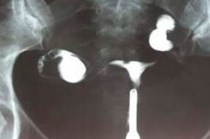
Ultrasound, X-ray examination, hysterosalpingography indicate insufficient organ size, incorrect configuration (tortuosity) of the fallopian tubes, small ovaries. The level of sex hormones (follicle-stimulating hormone, progesterone, estradiol, prolactin, luteinizing hormone, testosterone) and thyroid hormones (thyroid-stimulating hormone, T4) are necessarily examined. Many patients undergo uterine sounding, determination of bone age, X-ray of the sella turcica, magnetic resonance imaging of the brain. [ 13 ]
Additionally, a consultation with a therapist is required if extragenital pathologies are present, as well as consultations with an endocrinologist, urologist, surgeon, if there are disorders on the part of other related organs and systems.
Ultrasound for uterine hypoplasia is considered one of the most informative diagnostic studies. The procedure is performed using a vaginal and transabdominal sensor, longitudinal and transverse scanning. [ 14 ]
- Before a transabdominal gynecological ultrasound examination, the patient is prepared: an hour before the procedure, she should drink at least 1 liter of still water and not urinate until the end of the examination.
- Transvaginal ultrasound does not require any special preparation, and it is better to empty the bladder before the procedure.
Only a doctor can interpret the results of ultrasound diagnostics.
Echo signs of uterine hypoplasia are as follows:
- the organ length parameters do not correspond to the age and physiological norm;
- the cervix is large in size relative to the body of the uterus;
- a pronounced forward bend of the organ is noted;
- The fallopian tubes are thin, convoluted, and elongated.
The body of the uterus is normally slightly tilted forward, which is defined by such terms as "anteversio" and "anteflexio". The dimensions of the uterus are determined as transverse, longitudinal and anteroposterior indicators:
- the longitudinal indicator characterizes the length of the organ and is normally from 45 to 50 mm (in a woman who has given birth it can increase to 70 mm) + the length of the cervix should be 40-50 mm;
- the transverse indicator characterizes the width of the organ and is normally from 35 to 50 mm (in a woman who has given birth, it can increase to 60 mm);
- The anterior-posterior index indicates the thickness of the uterus and is normally between 30 and 45 mm.
The thickness of the endometrium varies throughout the monthly cycle. On the 5th-7th day of menstruation, its thickness is determined to be 6-9 mm. [ 15 ]
Often, only ultrasound is enough to diagnose uterine hypoplasia. Other studies are carried out to clarify the diagnosis and find the causes of the pathology, which is necessary for further correct and effective treatment.
Differential diagnosis
Type of pathology |
Quality of the monthly cycle |
Ultrasound signs |
Gynecological examination |
Anomalies of sexual development |
There is no menstrual function during puberty |
Signs of anomalies are detected: the cervix and body of the uterus are absent, there is a rudimentary horn or intrauterine septum, or a bicornuate uterus |
Signs of abnormal development of reproductive organs are detected |
Adenomyosis |
The monthly cycle is disturbed, menstrual bleeding is scanty or heavy, there is brown vaginal discharge, menstruation is painful |
The anteroposterior size of the uterus is increased, there are areas of high echogenicity of the myometrium, minor round anechoic formations (3-5 mm) |
The uterus is moderately painful, has nodes (endometriomas), and is enlarged |
Dysmenorrhea |
The monthly cycle is present, but patients complain of severe pain |
Typical echo signs are absent. |
No pathological signs are detected during gynecological examination. |
Inflammatory diseases of the pelvic organs |
Irregular, prolonged uterine bleeding |
Irregular uterine size and endometrial thickness, high degree of vascularization, fluid in the pelvis, thickened fallopian tubes, non-uniform decrease in echogenicity of myometrial zones |
Painfulness and softness of the uterus, presence of tubo-ovarian formations, intoxication symptoms |
Who to contact?
Treatment uterine hypoplasia
Treatment for uterine hypoplasia is prescribed taking into account the degree of pathology and pursues the following goals:
- elimination of the disorder, correction of organ parameters;
- restoration of the menstrual cycle, sexual and reproductive function;
- optimizing quality of life.
The basis of therapy for uterine hypoplasia is the use of replacement or stimulating hormonal drugs. Correctly selected treatment allows for an increase in the size of the organ sufficient for its normal physiological functions.
Additionally, physiotherapy treatment is used in the form of magnetotherapeutic, laser-therapeutic, diathermic, inductothermic, UHF procedures, balneotherapy, the use of ozokerite and paraffin. The basic goal of physiotherapy is to improve blood circulation in the uterine area.
An excellent effect is obtained from the endonasal galvanization procedure: this method involves stimulation of the hypothalamus-pituitary zone, which leads to increased production of hormonal substances, namely luteinizing hormone and follicle-stimulating hormone. [ 16 ]
To support and accelerate recovery, patients with uterine hypoplasia are recommended to take vitamin therapy, exercise therapy, manual therapy with gynecological massage, and spa treatment.
Vitamin complex preparations containing vitamins A, B, D, tocopherol, ascorbic and folic acids are used. Vitamin E has an antioxidant effect, stabilizes the monthly cycle, optimizes reproductive function. Vitamin C strengthens the vascular network, improves blood flow.
To improve reproductive function, a woman's diet should be reviewed. The doctor will definitely cancel strict diets and fasting, recommend sticking to a full diet, eating more fiber, vegetables and fruits, vegetable oils, cereals. Products such as spinach, broccoli and Brussels sprouts, tomatoes, sesame and flaxseed oil, seafood are especially recommended.
Medicines
Drug therapy is usually complex, including the use of drugs with different mechanisms of action.
- Hormonal agents:
- continuous course of estrogens during puberty;
- estrogens for the first phase of the monthly cycle, gestagens for the second phase.
In case of insufficient general somatic development, thyroid hormones are used (sodium levothyroxine 100-150 mcg per day), anabolic steroid drugs (methandrostenolone 5 mg 1-2 times per day, depending on the type of disorder). [ 17 ]
- Antibiotics are prescribed for frequent infectious processes:
- sulbactam/ampicillin (intravenous 1.5 g);
- clavulanate/ampicillin (intravenous 1.2 g);
- cefazolin (intravenously 2 g);
- cefuroxime (intravenously 1.5 g);
- vancomycin (if there is an allergy to beta-lactam antibiotics) 7.5 mg/kg every 6 hours or 15 mg/kg every 12 hours, for 7-10 days;
- ciprofloxacin 200 mg intravenously 2 times a day for a week;
- macrolide antibiotic azithromycin 500 mg once a day intravenously for 3-5 days.
Long-term hormonal therapy is often accompanied by undesirable side effects that all patients should be aware of:
- pain, enlargement of the mammary glands;
- increased appetite, sometimes nausea;
- dry mucous membranes;
- feeling of fatigue, weakness;
- thrombosis, thromboembolism.
It is important to understand that side effects do not occur in all patients, and their severity is also not the same. At the same time, without hormonal therapy, it is often impossible to correct the condition of the uterus and get rid of hypoplasia, since the growth and development of the organ directly depend on the production of hormones in the body.
Treatment with hormonal drugs
Hormonal drugs for uterine hypoplasia almost always become the main link in treatment. They help to balance the hormonal background, which helps to restore the development of the uterus.
Most often, the drugs of choice are the following hormonal agents:
- Femoston is a drug of estradiol and dydrogesterone, which activates the development of the entire reproductive system as a whole, including the fallopian tubes. The treatment is long-term, with breaks: the scheme is drawn up by the attending physician, taking into account the individual characteristics of the patient and the reaction of her body to the treatment.
- Duphaston is very often prescribed for uterine hypoplasia. This hormonal agent is an artificial analogue of progesterone, which is especially relevant when it comes to endometrial hypoplasia. Duphaston stabilizes the balance of hormones in the body if taken in combination with other complex drugs. The duration of treatment is usually more than six months. The dose and regimen are determined by the doctor individually.
- Estrofem is a drug that helps stabilize the balance of estrogens in the female body, activate the development of the main reproductive organ, and improve the function of the fallopian tubes. At the same time, the monthly cycle is established. Take 1 tablet daily in the morning. The duration of the treatment course is determined by the doctor individually. As a rule, the courses are short-term (about 2 months), after which you need to take a break.
- Ovestin contains a natural female hormone - estriol. This hormone interacts with the nuclei of endometrial cells, normalizes the condition of the epithelium. As a rule, the drug is used in the form of suppositories: 1 suppository is inserted per day with a slow decrease in dosage, depending on the dynamics of treatment. Vaginal suppositories are inserted into the vagina in the evening, before going to bed.
- Microfollin is an ethinyl estradiol preparation that eliminates disorders associated with endogenous estrogen deficiency, stimulates proliferation of the endometrium and vaginal epithelium, and promotes the development of the uterus and secondary sexual characteristics of women with hypoplasia.
Hormonal treatment should never be carried out independently: such drugs are always prescribed by a doctor, and then their intake is monitored, adjusting the dosage and frequency of use. The reaction of the woman's body to hormonal therapy and the dynamics of treatment must be taken into account. [ 18 ]
Physiotherapy treatment
Physiotherapy procedures are successfully used as an addition to the doctor's main prescriptions for uterine hypoplasia. The following are especially common:
- Magnetic therapy, using a magnetic field, has an anti-edematous and anti-inflammatory effect, improves blood circulation and stimulates cellular structures.
- Ultrasound therapy affects the organ at the cellular level, stimulates tissue metabolism, which is combined with pronounced heat production. When the temperature rises, blood circulation improves, pain syndrome disappears, and adhesions soften. In addition, ultrasound vibrations activate the hormonal function of the ovaries, which helps to establish the monthly cycle.
- Phonophoresis allows to deliver drugs directly to the pathological focus using ultrasound waves. This allows the drug to act locally, which significantly reduces the likelihood of side effects. Most often, antibacterial drugs, anti-inflammatory and vitamin agents are delivered to tissues by phonophoresis.
- Electrophoresis “works” in a similar way to phonophoresis, but electric current is used to deliver medications.
In addition, with uterine hypoplasia, gynecological massage sessions are indicated: 10 minutes daily for 1-1.5 months. Gynecological vibration massage optimizes lymph and blood circulation in the pelvis, which eliminates congestion and enhances metabolic processes. Thanks to vibration massage, it is possible to strengthen the ligamentous-muscular system of the uterine organ and pelvic floor. Inductothermy and acupuncture are also useful. [ 19 ]
Herbal treatment
Traditional methods of therapy for uterine hypoplasia can be used, but they will have a real beneficial effect only in combination with the main drug treatment. In other words, full-fledged conservative treatment cannot be replaced by home remedies, but it is quite possible to supplement it.
Herbal teas, decoctions and infusions based on herbs that have anti-inflammatory and hormonal activity are successfully used as herbal remedies to correct uterine hypoplasia.
- The orthilia secunda, or one-sided wintergreen, contains both phytoestrogens and plant progesterone, so the plant is indicated for the treatment of many gynecological diseases. Most often, one-sided wintergreen tincture is used at home. To prepare it, take 100 g of dry crushed plant, pour 1 liter of vodka, place in a dark place. Keep under the lid for 2 weeks, then filter and begin taking: 35 drops with a small amount of water between meals, twice a day. Treatment is usually long-term, several months. The drug should not be taken in childhood.
- Knotweed, or bird's highlander, has anti-inflammatory, antibacterial, diuretic, antitumor, and analgesic effects. Due to the phytonutrients included in the plant, knotweed can stimulate the female reproductive system, increase hormone production, and stabilize the monthly cycle. The plant is taken in the form of a decoction. Dried grass in the amount of 20 g is poured with 200 ml of boiling water, infused for an hour under a lid. Take one sip 3-4 times a day 30 minutes before meals.
- Sage can stimulate the production of estrogens by the female body and regulate ovulation. Preparations from the plant are taken in the first phase of the cycle, after the end of menstrual bleeding (approximately on the 4th-5th day). Sage should not be taken for endometriosis, tumors, or severe hypertension. To prepare the medicine, take 1 tbsp. of the dry plant, brew 200 ml of boiling water, leave until cool, strain and store in the refrigerator. During the day, you need to drink the entire infusion, which is approximately 50 ml 4 times a day.
- Elecampane successfully stabilizes the menstrual cycle, improves blood circulation in the periuterine area, thereby promoting the development of the organ. To prepare an infusion of the plant, 2 tablespoons of raw material are poured with 0.5 liters of boiling water and kept under a lid for half an hour. Then the infusion is filtered and divided into two halves: one part is drunk in the morning half an hour before breakfast, and the second - an hour before dinner. Take the medicine daily. If nausea or weakness occurs, the dosage is reduced.
- Red brush is a natural herbal remedy that is actively used to treat myomas, fibroids, mastopathy, cervical erosions, polycystic ovary disease, irregular menstruation and even uterine hypoplasia. To treat hypoplasia, use a tincture of the plant: 50 g of dry crushed raw materials are poured with 0.5 liters of vodka, infused in a sealed form in a dark place for one month (sometimes you need to shake it). Then filter the tincture and start taking 1 teaspoon three times a day 40 minutes before meals. The treatment regimen is as follows: four weeks of intake - two weeks of break.
Surgical treatment
In case of concomitant endometrial hypoplasia against the background of the lack of effectiveness of conservative therapy, the doctor may prescribe surgical intervention, which involves separate diagnostic curettage. The operation consists of resection of the inner uterine layer (the so-called cleaning) to activate the processes of renewal and subsequent growth of the functional layer of the endometrium.
The intervention is performed using general intravenous anesthesia through vaginal access (without incisions).
The execution of surgical manipulations is controlled through a hysteroscope, making the operation precise and safe.
The surgical intervention lasts up to half an hour, after which the patient is placed in a day hospital ward, where she is under the supervision of medical specialists for several hours. If she feels well and there are no complications, the woman can go home that same day. [ 20 ]
Prevention
Preventive measures are necessary, first of all, during preparation for pregnancy and at the stage of conception. Primary prevention of uterine hypoplasia may include the following measures:
- Proper nutrition for women during the reproductive period, providing the female body with all the necessary vitamins and microelements, taking dietary supplements recommended by the doctor.
- Avoid smoking and drinking alcohol, both at the planning stage and during pregnancy. You should also beware of harmful foods and drinks.
- Preventing exposure of the female body to hazardous substances, in particular heavy metals, pesticides, and certain medications.
- Timely prevention of infectious diseases, vaccination (for example, the rubella vaccine can be administered at least 4 weeks before pregnancy to those patients who have not been previously vaccinated and have not had rubella in childhood).
It is necessary to take care of the health of the girl's entire reproductive system from the moment of her birth. It is advisable to show the child to a doctor - a pediatric gynecologist - already in infancy. This is necessary so that the specialist can assess the development of the baby's genitals.
Both in early childhood and at subsequent age stages, the child should be protected from stress, provided with normal nutrition, maintain hygiene, and prevent infectious and inflammatory diseases.
In a very important age period – adolescence, starting from about 11 years old, a girl needs to be especially carefully protected from infectious pathologies, and especially from viral ones. It is necessary to eliminate all possible sources of infection in the body – for example, caries, chronic tonsillitis, etc.
Explanatory work with children plays a very important role: it is important to explain to the child why smoking, drinking alcohol, using drugs and toxic substances are harmful. These factors cause great harm to the child's body, since they have gonadotoxicity.
Regular lack of sleep, fasting, early onset of sexual activity, and psycho-emotional overload have a negative impact on the development of the female reproductive system as a whole.
Forecast
If uterine hypoplasia is caused by disorders of the endocrine system, then timely treatment can be effective. However, a severe form of congenital defect cannot be corrected, and the probability of a woman becoming pregnant is practically reduced to zero. [ 21 ]
Treatment for a relatively small degree of hypoplasia is long-term, but the prognosis is often favorable: many women manage to successfully carry and give birth to a long-awaited baby.
It is important to understand that patients must immediately prepare for long-term therapy with strict adherence to all medical prescriptions. The outcome of this treatment depends on the degree of the anomaly and the reasons for its occurrence. Hypoplasia of the uterus is not always completely cured. However, doctors often manage to achieve the main desired result: women become pregnant and become mothers. The main thing is to find a good specialist who will competently select an approach to treatment.