Medical expert of the article

New publications
Ribs
Last reviewed: 06.07.2025

All iLive content is medically reviewed or fact checked to ensure as much factual accuracy as possible.
We have strict sourcing guidelines and only link to reputable media sites, academic research institutions and, whenever possible, medically peer reviewed studies. Note that the numbers in parentheses ([1], [2], etc.) are clickable links to these studies.
If you feel that any of our content is inaccurate, out-of-date, or otherwise questionable, please select it and press Ctrl + Enter.
Twelve pairs of ribs and the sternum, together with the thoracic spine, form the rib cage.
The ribs (costae) are long, narrow, thin, curved bony plates. In front, the bony part of the rib continues into the cartilaginous part - the costal cartilage (cartilago costalis). The seven upper pairs of ribs, connected in front with the sternum, are called true ribs (costae verae). Ribs VII, IX and X are connected with their cartilages to the cartilaginous part of the rib above - these are false ribs (costae spuriae). Ribs XI and XII end in the thickness of the abdominal muscles. These ribs are called fluctuating (costae fluctuantes).
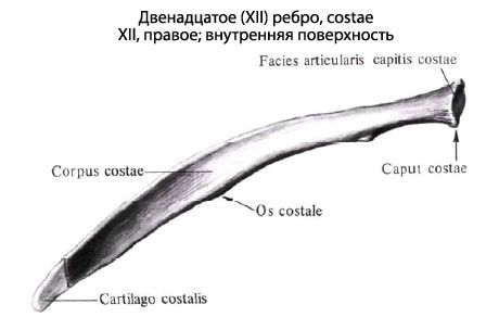
At the posterior end of each rib there is a thickening - the head of the rib (caput costae), which connects with the corresponding costal fossa on the thoracic vertebrae. On the head of the II-X ribs there is a crest of the head of the rib (crista capitis costae), since each of these ribs connects with two costal fossae. The heads of the XI and XII ribs do not have a crest.
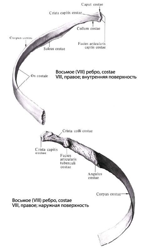
Anterior to the head of the rib is the narrow neck of the rib (collum costae), which passes into its body (corpus costae). At the 1st-10th ribs, at the border of the neck and body, there is a tubercle (tuberculum costae) with an articular surface for articulation with the transverse process of the corresponding vertebra. The flattened body of the rib has a convex outer and concave inner surface. On the inner surface, at the bottom along the rib, there is a costal groove (sulcus costae), to which the intercostal vessels and nerve are adjacent. Slightly lateral to the tubercle is the rounded angle of the rib (angulus costae).
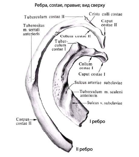
The first rib differs from the other ribs. It has an upper and lower surface, a lateral and medial edge. Not far from the junction with the sternum on the upper surface is the tubercle of the anterior scalene muscle (tuberculum musculi scaleni anterioris). In front of the tubercle is the groove of the subclavian vein (sulcus venae subclaviae), and behind the tubercle is the groove of the subclavian artery (sulcus arteriae subclaviae).
What do need to examine?
How to examine?