Medical expert of the article

New publications
Female genital prolapse
Last reviewed: 07.07.2025

All iLive content is medically reviewed or fact checked to ensure as much factual accuracy as possible.
We have strict sourcing guidelines and only link to reputable media sites, academic research institutions and, whenever possible, medically peer reviewed studies. Note that the numbers in parentheses ([1], [2], etc.) are clickable links to these studies.
If you feel that any of our content is inaccurate, out-of-date, or otherwise questionable, please select it and press Ctrl + Enter.
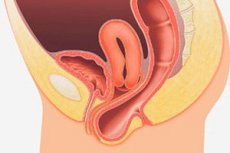
Prolapse of the genitals is a polyetiological disease, the basis of which is dystrophy and failure of the ligamentous apparatus of the uterus and pelvic floor muscles, increased intra-abdominal pressure. Pelvic structures: uterus (uterine prolapse) or vagina (vaginal prolapse), anterior vaginal wall (bladder hernia), or posterior vaginal wall (rectocele).
 [
[ Risk factors
Factors that contribute to the development of pelvic floor muscle failure include pathological childbirth, estrogen deficiency, age-related changes in muscle and connective tissue, genetic predisposition, as well as a number of extragenital diseases and unfavorable social conditions.
Pathogenesis
In the pathogenesis of prolapse and/or genital prolapse in young nulliparous women (or those with only uncomplicated births) with unchanged hormonal background and normal social conditions, the leading role is played by a systemic defect of connective tissue. Under the influence of any of the listed factors or their combined effect, functional failure of the ligamentous apparatus of the internal genital organs and the pelvic floor occurs. Against the background of functional failure of the ligamentous apparatus of the uterus and its appendages and increased intra-abdominal pressure, the organs begin to extend beyond the pelvic floor. In this case, several variants of pathogenetic mechanisms of uterine and vaginal prolapse are distinguished:
- the uterus is located entirely inside the extremely expanded single fundus; deprived of any support, it is squeezed out through the pelvic floor;
- part of the uterus is located inside, and part is outside the hernial orifice; the first part is squeezed out, while the other is pressed against the supporting base.
In the second variant, the vaginal part of the cervix, due to the constant pressure inside the hernial orifice, can descend and stretch (elongatio coli); while the body of the uterus, lying outside the hernial orifice and adjacent to the still partially functioning levator ani, resists complete prolapse of the organ. This mechanism explains the formation of an elongated and thinned uterus, the elongation of which depends exclusively or mainly on the hypertrophy of the cervix, while the fundus of the uterus may remain in an almost correct position at this time. In such a situation, complete prolapse of the uterus occurs with its retroflexion - when the axis of the uterus coincides with the axis of the vagina. Therefore, retroflexion is considered a risk factor for complete prolapse of the uterus.
In clinical practice, the classification of prolapse of the female genital organs proposed by K. F. Slavyansky is still used.
Symptoms female genital prolapse
The most common complaints of patients with prolapse of the internal genital organs are: aching pain and/or a feeling of heaviness in the lower abdomen, leucorrhoea, sexual dysfunction, a feeling of a foreign body in the vagina, urinary and gas incontinence during physical exertion, coughing, sneezing.
Stages
Classification of downward displacements of the vagina (according to K. F. Slavyansky):
- 1st degree. Prolapse of the anterior vaginal wall, the posterior wall, or both together (the walls do not extend beyond the entrance to the vagina).
- 2nd degree. Prolapse of the anterior or posterior vaginal walls, as well as both together (the walls are located outside the vaginal opening).
- 3rd degree. Complete prolapse of the vagina, which is accompanied by prolapse of the uterus.
What do need to examine?
Who to contact?
Treatment female genital prolapse
The anatomical and topographic features of the pelvic organs, common blood supply, innervation, and close functional connections allow us to consider them as a single system in which even local changes cause damage to the function and anatomy of neighboring organs. Therefore, the main goal of prolapse treatment is to eliminate not only the underlying disease, but also to correct disorders of the genitals, bladder, urethra, rectum, and pelvic floor.
Among the factors that determine the treatment tactics for patients with genital prolapse, the following are distinguished:
- degree of prolapse of the genitals;
- anatomical and functional changes in the genital organs (the presence and nature of concomitant gynecological diseases);
- the possibility and feasibility of preserving and restoring reproductive and menstrual functions;
- features of dysfunction of the colon and sphincter of the rectum;
- age of patients;
- concomitant extragenital pathology and the degree of risk of surgical intervention and anesthetic care.
General strengthening treatment. This type of therapy is aimed at increasing tissue tone and eliminating the causes that contribute to the displacement of the genitals. Recommended: proper nutrition, water procedures, gymnastic exercises, changing working conditions, uterine massage.
Surgical treatment of prolapse of genital organs. Pathogenetically justified method of treatment of prolapse of female genital organs should be considered surgical intervention.
To date, over 300 methods of surgical correction of this pathology are known.
Known methods of surgical correction of genital prolapse can be divided into 7 groups, based on the anatomical structures that are strengthened to correct the incorrect position of the genitals.
- 1st group of operations – strengthening of the pelvic floor – colpoperineolevatoroplasty. Considering that the pelvic floor muscles are always pathogenetically involved in the pathological process, colpoperineolevatoroplasty should be performed in all cases of surgical intervention as an additional or primary aid.
- The 2nd group of operations – the use of various modifications of shortening and strengthening of the round ligaments of the uterus. The most frequently used is shortening of the round ligaments with their fixation to the anterior surface of the uterus. Shortening of the round ligaments of the uterus with their fixation to the posterior surface of the uterus, ventrofixation of the uterus according to Kocher and other similar operations are ineffective, since the round ligaments of the uterus, which have high elasticity, are used as a fixing material.
- The 3rd group of operations is strengthening the fixing apparatus of the uterus (cardinal, uterosacral ligaments) by suturing them together, transposition, etc. This group includes the “Manchester operation”, the essence of which is to shorten the cardinal ligaments.
- The 4th group of operations is rigid fixation of prolapsed organs to the walls of the pelvis - to the pubic bones, sacrum, sacrospinal ligament, etc. Complications of these operations are osteomyelitis, persistent pain, as well as so-called operative-pathological positions of the pelvic organs with all the ensuing consequences.
- The 5th group of operations is the use of alloplastic materials to strengthen the ligamentous apparatus of the uterus and fix it. The use of these operations often leads to rejection of the alloplast and the formation of fistulas.
- The 6th group of operations is partial obliteration of the vagina (median colporrhaphy according to Neugebauer-Lefort, vaginal-perineal cleisis - Labhardt operation). The operations are non-physiological, exclude the possibility of sexual activity, and relapses of the disease are observed.
- The 7th group of operations is a radical surgical intervention - vaginal hysterectomy. Of course, this operation completely eliminates the organ prolapse, however, it has a number of negative aspects: relapses of the disease in the form of enterocele, persistent disruption of menstrual and reproductive functions.
In recent years, the tactic of combined correction of genital prolapse using laparoscopy and vaginal access has become popular.
Orthopedic methods of treating prolapse of the genitals. Methods of treating prolapse and prolapse of the genitals in women using pessaries are used in old age when there are contraindications to surgical treatment.
Physiotherapeutic treatment. Timely and correctly applied methods of physiotherapy and diadynamic sphincterotonization are of great importance in the treatment of pubic genitalia and urinary incontinence in women.