Medical expert of the article

New publications
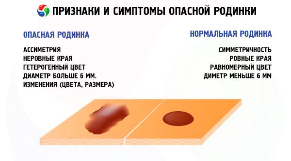
A pink mole
Last reviewed: 04.07.2025

All iLive content is medically reviewed or fact checked to ensure as much factual accuracy as possible.
We have strict sourcing guidelines and only link to reputable media sites, academic research institutions and, whenever possible, medically peer reviewed studies. Note that the numbers in parentheses ([1], [2], etc.) are clickable links to these studies.
If you feel that any of our content is inaccurate, out-of-date, or otherwise questionable, please select it and press Ctrl + Enter.
Causes pink mole
A pink mole occurs as a result of a malfunction in the capillaries and vessels that supply blood to the skin. This mole contains many microscopic vessels and can appear on any part of the human body. They are found mainly in teenagers or children - this is probably due to the fact that it is in childhood that the circulatory system undergoes some changes.
Such moles can also appear in adults as a result of prolonged exposure to sunlight. They can grow in various layers of the skin and parts of the circulatory system - capillary, arterial, venous.
The reasons for the appearance of pink moles may be hormonal changes that have begun in the body. Some experts also believe that their appearance may be the result of a gastrointestinal disease (often the pancreas).
Another reason is excessively intense functional activity of blood vessels, or a deviation in the activity of pigment cells that perform the process of skin pigmentation.
Thus, if you notice a pink mole on your skin, you can visit a doctor for preventive purposes - this will help avoid possible complications.
Pathogenesis
Moles that are pink in color are also called angiomas, and they are considered completely benign. They are of vascular origin and mainly appear in children. Although pink moles often grow in adults.
Doctors are not too concerned about this phenomenon, because such moles are not at all dangerous for the body, and also do not affect either its biological processes or general functional activity.
If we talk about children, the pathogenesis of their pink birthmarks is quite specific - they can appear and disappear regardless of external factors and any impact.
Pink nevi can grow in any layer of the skin - they appear on capillary, venous, arterial areas. These moles will differ in appearance, depending on where exactly they are located. Therefore, they also need to be treated in completely different ways.
Typically, moles of this type appear as a result of a disorder in the capillary vascular section. They arise from the cells of the vessels and grow inside the skin layer.
On the surface, a person sees them as small, convex pink moles or small red formations. Among congenital benign tumors, such types of angiomas are considered the most common.
Symptoms pink mole
A pink mole is formed on the skin from capillaries. It is a benign formation that does not pose a threat to life. Among its main symptoms are the following signs:
- Red spots that form on the skin right from the moment of birth or appear later in life;
- A reddish rash that makes the skin look like blood vessel markings;
- With their appearance, the patient's health does not worsen, and the temperature does not rise.
Simple pink moles often form on the body unnoticed by a person, they do not affect the body in any way and do not worsen the state of health. If you have any concerns about the condition of the mole, you need to go to see a doctor - he will be able to make an accurate diagnosis.
Pink raised mole
A pink mole is also called an angioma. It is a benign tumor that forms from blood vessels (capillaries). Such moles are very common on the body - about 22% of all nevi on the skin are pink or red moles.
These reddish spots appear due to a congenital malformation of the blood vessels. They appear in babies, from the moment of birth.
These nevi can have a variety of shapes – they can be completely flat or slightly convex. The sizes can also vary greatly – there are very small pinkish or red dots, and huge spots that can even occupy the entire arm or leg.
Simple, or as they are also called capillary, moles are mainly pink, purple or crimson spots. When pressed, they turn pale. These moles can be convex and smooth. Some of them look like a formation with a red dot in the center, from which small dilated vessels radiate.
A pink, raised mole usually appears in mature or older people. It indicates an early stage of skin cancer (usually squamous cell carcinoma or basalioma).
The mole turned pink
Moles turn red or swell when injured. They can also change color due to various diseases.
What should you do if a mole turns pink? If a mole changes color, you should definitely see a doctor, as well as if it has changed shape or started to cause you pain and discomfort. At the clinic, you can undergo a special examination that will help make sure that the formation does not contain malignant cells and find out whether it needs to be removed from the body.
It is not recommended to solve the problem of a pink mole on your own - you should not use folk remedies or try to remove it yourself. Such home "treatment" can lead to at least an infection, which will result in inflammation. In the worst case, you will face the development of a pathological focus, which will take a very long time to treat, spending a lot of effort on it.
A pink mole that is very painful and bleeding can be removed using a radio knife or laser. It can also be cauterized. A qualified doctor will prescribe the appropriate treatment based on the examination data and additional diagnostics.
 [ 8 ]
[ 8 ]
Complications and consequences
A pink mole in itself is not considered a malignant formation, but it should be understood that if it is damaged (this is especially easy to do if it has a convex shape), various consequences and complications may arise, up to the development of melanoma.
Therefore, in case of any damage, you should contact a doctor to have it removed. The removal procedure is completely safe and does not entail any negative consequences.
Diagnostics pink mole
If you have never had all of your moles examined before, it is recommended that you see a qualified dermatologist.
During the diagnostics, all moles will be examined, as dangerous formations can be on any part of your body. It is necessary to make sure that the surface of the feet and palms, as well as the areas between the fingers, behind the ears, nails and mucous membranes, skin folds have been carefully checked. Most of all, doctors are often interested in those moles that are very different from all the others.
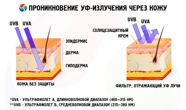
Preventive examinations should be taken by those who are often exposed to ultraviolet radiation:
- Frequent visitors to the solarium;
- Those undergoing a course of treatment that uses ultraviolet rays;
- Before and after a vacation in a hot southern country.
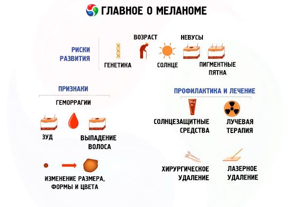
There are several risk groups that are more likely to develop melanoma. These people should undergo examinations as often as possible:
- blood relatives of people diagnosed with melanoma;
- fair-skinned people who are very sensitive to ultraviolet rays;
- people who have a lot of moles.
As soon as you notice that your pink mole or any other has changed, or a new nevus has appeared, you should immediately consult a doctor. And it is not recommended to delay the visit.
[ 11 ]
Tests
If your pink mole has started to bother you, you should visit a doctor who will determine the reason for its change. In some cases, it may be necessary to remove the nevus and send it for histological analysis to find out whether there are malignant cells in the formation.
Instrumental diagnostics
Even an experienced doctor sometimes cannot detect the early stage of a mole's transformation into a malignant tumor. That is why a biopsy is sometimes performed to confirm the diagnosis, after which the removed tissue is sent for histological analysis.
There is also a method using Computer Epiluminescent Dermatoscopy – it is the newest method of instrumental diagnostics of degenerating nevi. This latest technology allows dermatologists to examine formations not only on the surface of the skin, but also to discern any changes that occur in deeper layers.
Thanks to this procedure, it is possible to see the processes occurring deep in the nevus without damaging the tissue. The data obtained during this diagnostics will be analyzed by a doctor, after which it will become clear what the degree of risk is due to the transformation of the mole. The doctor will also give recommendations on how to behave with it in the future or refer the patient for an operation to remove it.
Currently, the dermatoscopy method is considered the most reliable. It allows diagnosing malignant degeneration of a nevus at an early stage. It also makes it possible to clarify whether the patient's pink mole is malignant at all.
Differential diagnosis
Differential diagnostics of moles is carried out to find out whether they contain malignant cells. To do this, they are removed from the body, after which they are sent for a biopsy - this study will give a comprehensive answer and help make the correct diagnosis. A pink mole can also be examined using a dermatoscope - this device helps make a diagnosis, detecting a malignant formation at an early stage.
Who to contact?
Treatment pink mole
There are several methods for removing a pink mole. It should be noted right away that cauterization is not a good method in this case, since moles are usually located deep under the skin, and only the upper part protrudes to the surface. Therefore, after such removal, the roots of the nevus will remain in the skin, which is why it may appear in the same place again after some time.
The most appropriate method of removal is chosen by the doctor after examining the nevus. He will also determine whether there are malignant formations in it.
Usually, mole removal is performed using a laser. Among the most modern procedures are infrared or light coagulation of vessels, X-ray treatment and sclerotherapy of the vascular bed. Nevi with a flat structure are easier to remove than convex ones. During the process of nevus removal, if necessary, anesthetic cream can be used, but anesthesia is generally not used.
Remember that the removal of a mole itself can be an unpleasant operation, after which small reddish spots may remain on the skin, although they disappear completely after some time. After the pink mole is removed, you should avoid going to a solarium for at least 1 month and spend less time in the sun.
Medicines
A pink mole is not considered a health hazard, so it does not need to be treated. Medicines are not used in this case.
Folk remedies
Moles can be removed using folk remedies, although it should be understood that such methods cannot be considered completely safe.
Every day before going to bed, lubricate the mole with 1 drop of vinegar essence.
Grind several garlic cloves into a mush and mix them with 1 tbsp. butter and 50 g honey. As a result, you will get an ointment that you need to smear the nevus with. Wash off the ointment with warm water after 4 hours. The course of treatment lasts 1 month.
1-2 times a day, apply celandine juice to the nevus.
Make an ointment from 1 tbsp of dandelion root juice and 4 tbsp of butter, which you should apply to the mole twice or three times a day.
Remove the kernels from the cherry pits (100g), grind them into powder. It should be poured with half a liter of olive oil and the resulting mixture should be infused for 2 weeks in the dark. The ointment should be applied to the nevus every day, kept for 20 minutes, and then washed off with warm water.
Mix willow ash with vinegar and apply the resulting mixture to nevi 2-3 times a day.
A pink mole can be removed using an ointment made from 8 tablespoons of hemp oil and 2 tablespoons of crushed chalk. The mixture should be left to infuse for 1 week. The mole should be smeared 2-3 times a day for a month.
Apply fresh onion juice to the mole several times daily.
Apply pineapple juice to nevi 3-4 times a day.
2 cut garlic cloves insist for 2 weeks in apple cider vinegar (half a glass). The tincture is applied to the mole, having previously soaked a piece of cotton cloth in it. This procedure should be done until the nevus completely disappears.
Make a mixture of castor/linseed oil and honey (equal halves) and apply it to the nevus, leave for a few minutes, then wash off. The procedure should be performed three times a day.
[ 14 ]
Herbal treatment
A pink mole can be removed through herbal treatment. This is also not the most reliable and safe method, but if you have no other options, you can use it.
Every night until the mole disappears, attach a fresh crushed calendula flower to it.
Grind rosehip petals and apply the resulting powder to the nevus twice/three times daily until it disappears.
Take chopped celandine and Vaseline (equal proportions), mix, and apply the resulting ointment to the mole every day.
Prevention
To prevent a situation where your pink mole degenerates into a malignant skin formation, and to protect yourself from the possibility of developing melanoma, you should take preventive measures. Try to follow the following rules:
- Limit your time in the sun as much as possible (this applies especially to the summer period and lunchtimes);
- If you have to be in the sun anyway, protect your skin from the sun's rays by wearing a wide-brimmed hat, a long-sleeved shirt and trousers;
- If you have to be in direct sunlight, use sunscreen with a protection factor of at least 15;
- Try to examine the surface of your skin as often as possible, checking for old moles and looking for possible new ones;
- Find out what the primary and secondary symptoms of melanoma are, and consult your doctor about this. You should understand what the external signs of melanoma are and what distinguishes it from a simple benign mole.
If you have suspicions about any nevus, you should immediately contact a dermatologist, because the sooner you detect the development of melanoma, the higher the chance of its successful treatment.
Forecast
In some cases, a pink mole can degenerate into a malignant formation – superficial basalioma or melanoma.
Such superficial basaliomas look like a slightly flaky reddish-brownish plaque with a shiny surface and raised edges. They are located mainly on the body and usually appear in multiple quantities at once. The prognosis for the course of such a disease is quite optimistic; it can exist for decades, gradually increasing its area.