Medical expert of the article

New publications
MRI of the knee: what it shows, deciphering the results
Last reviewed: 03.07.2025

All iLive content is medically reviewed or fact checked to ensure as much factual accuracy as possible.
We have strict sourcing guidelines and only link to reputable media sites, academic research institutions and, whenever possible, medically peer reviewed studies. Note that the numbers in parentheses ([1], [2], etc.) are clickable links to these studies.
If you feel that any of our content is inaccurate, out-of-date, or otherwise questionable, please select it and press Ctrl + Enter.
The knee is one of the joints that is most often subject to various injuries and damages, both in childhood and in the elderly. Fortunately, most often the injury is limited to abrasions and hematomas. But sometimes the pain does not go away for quite a long time, or even intensifies, in such a situation the doctor may prescribe an MRI of the knee joint to diagnose the pathological condition.
MRI of the knee joint will always help the doctor to determine the existence of a problem and prescribe adequate treatment. This type of examination is completely safe, and in terms of information content it surpasses most other similar methods.
Indications for the procedure
In case of problems with the knee joint, doctors are in no hurry to prescribe procedures such as MRI - this type of examination is quite expensive. Therefore, such diagnostics are carried out only if there are compelling indications, which include:
- arthritis with a complex course, of infectious or rheumatoid origin;
- congenital defect of the knee joint;
- severe inflammatory process associated with a gout attack;
- collagen disease involving the knee ligaments;
- meniscopathy;
- complex traumatic injuries of the knee joint;
- tumor processes of primary or metastatic nature;
- gonarthrosis;
- chronic instability of the knee joint;
- unexplained cause of knee joint pain;
- ligament damage;
- inflammation of the knee joint capsule;
- preoperative and postoperative period associated with surgical intervention in the knee joint area.
It cannot be said that all the listed indications are absolute - in each specific case, the doctor decides everything. In case of milder pathologies and injuries, MRI can be easily replaced by radiography, but in complex cases, you cannot do without MRI.
- In case of injury, it will help to determine the location and extent of damage, stretching, rupture of meniscus, ligaments, muscles. This type of diagnostics is appropriate for fractures, cracks, as well as severe bruises with hemorrhages and bleeding.
- MRI of the knee joint with a meniscus rupture does not require the use of contrast: the meniscus on the image looks like a dark stripe, and all damage is clearly displayed in a white shade. A meniscus rupture can occur with sudden motor activity in the lateral direction, or in a direction that goes beyond the capabilities of the joint.
- In case of synovitis, it is performed due to the non-specificity of the symptoms of this disease. When synovitis occurs, the doctor who describes the MRI notes a change in the intensity of the signal in the joint cavity (due to the accumulation of fluid). This signal has increased intensity in the T2WI mode, and decreased intensity in the T1WI mode. If MRI is performed with the introduction of contrast, the signal from the affected membrane will be amplified. Growths hanging inside the folds of the joint capsule are visualized.
- In arthrosis and arthritis, it can be difficult due to a large accumulation of fluid - effusion. For accurate diagnostics, MRI of the knee joint is performed in the frontal projection.
- A cruciate ligament rupture most often shows no ligament imaging due to local edema and hemorrhage after injury. An incomplete rupture, which is visible as a widened cruciate ligament with a hyperintense signal and visible intact fibers, is called an interstitial rupture. This image must be differentiated from degenerative processes in the intact ligament.
How often can you have an MRI of the knee joint?
Magnetic resonance imaging is performed for various diseases of the knee joint. This type of diagnostics is performed as often as necessary. In most patients, the initial MRI helps the doctor confirm or refute the diagnosis and begin correct and effective treatment. Additional MRI procedures may be prescribed to clarify some previously questionable points in the diagnosis, as well as to assess the condition of the joint after surgery, to monitor the effectiveness of the therapy, for a more detailed study using contrast.
Electromagnetic radiation does not pose any threat of radiation load to the patient's body - this is its main difference from X-ray. Therefore, MRI is allowed to be performed as many times as necessary for adequate treatment. Specialists insist: MRI is safe and extremely informative.
Preparation
There is no need to prepare in advance for an MRI of the knee joint: you do not need to follow a special diet, take any medications or fast. The only requirement is to leave all metal items at home, including jewelry, watches, and other accessories.
During the procedure, the patient will have to take off some clothing: for example, during an MRI of the knee, these are trousers, tights, a skirt, etc.
Be sure to inform your doctor if you are pregnant, have any allergies to medications, have chronic illnesses, or have metal implants or pacemakers.
The device for carrying out the procedure
An MRI machine for a standard examination of the knee joint should have a power of 1.5 Tesla. If a more accurate image of the tissue structure is required, then a power of 1 Tesla can be selected - however, this type of machine is more in demand for diagnosing the brain and abdominal organs.
There are also varieties of closed and open type devices:
- closed type can have a power of 1-3 Tesla;
- open type (suitable for patients with claustrophobia) has a power of up to 0.4 Tesla.
The image is more informative if the magnetic power is higher, so doctors recommend choosing an MRI machine with 1.5 Tesla power ratings.
It is better, if possible, to choose a high-field device for MRI of the knee joint - that is, a closed type. It gives a better image than that obtained on open devices. It is especially important to conduct a high-quality study if it is necessary to visualize the ligament and tendon system.
Technique MRI of the knee joint
MRI of the knee joint is performed in almost the same way as MRI examination of other parts of the body. Diagnostics are carried out in stages:
- The patient lies horizontally on a special mobile couch, the doctor fixes his limbs and head using belts and/or pads for this purpose. This action is necessary to prevent the patient from accidentally making a movement that will subsequently affect the quality of the image.
- The mobile couch is placed inside the CT machine, and the doctor begins scanning, during which a constant noise is heard.
- For the patient's convenience, the internal chamber of the tomograph is equipped with lighting and a ventilation system, as well as a voice connection, through which the patient can maintain communication with the doctor.
- Once the examination is complete – approximately 15 minutes later – the patient leaves the machine and can return to their normal activities. Sometimes it may take some time to receive the MRI report if it is not sent directly to the attending physician.
How is an MRI of the knee joint performed?
- A conventional "closed" MRI machine looks like a volumetric cylindrical tube with a powerful magnet located around its circumference. During the diagnostics, the patient lies on a pull-out couch, which is moved into the center of the magnetic radiation at the beginning of the procedure. An "open" MRI has a similar operating principle, but in such a machine the magnet is not located around the circumference, but only on the sides of the patient.
Open MRI of the knee joint is suitable for those people who suffer from claustrophobia or obesity.
- MRI of the knee joint ligaments helps the doctor to examine the problem in different planes. This way, it is possible to evaluate not only the existing problem, but also to detect associated tissue damage, if any.
- MRI of the right and left knee joints is performed by placing special coils on the affected area. To obtain a correct image, it is necessary to ensure a motionless position of the body and limbs for about a quarter of an hour. If contrast is administered, the examination time may be extended. The patient should not experience any discomfort during the procedure. Sometimes there may be a feeling of warming of the knee - this is an adequate reaction of the tissues to the radiation of the magnet.
- MRI of the knee joint with contrast helps to see hemorrhages, bleeding, inflammatory foci, instability of blood supply, tumors. The essence of contrast is that special substances are injected into the patient's vein that can enhance magnetic resonance. The contrast component diverges through the blood vessels and settles in the tissues: the larger the vascular network in the organ being examined, the clearer the image becomes. In the area of hemorrhages or injuries, or in the presence of an inflammatory focus, the degree of blood flow will differ from that in healthy areas. In tumors, which have their own saturated capillary network, contrast is especially clear. Before performing contrast MRI, you need to make sure that the patient is not allergic to the injected substance. If there is no allergy, then the use of contrast is absolutely safe: the substance is eliminated from the body on its own within 1-2 days. Contrast should not be used if the patient suffers from renal failure or acute inflammation of the urinary tract.
How long does it take and what does an MRI of the knee joint show?
The MRI procedure of the knee joint takes 30 minutes. The MRI information reading itself takes about 15 minutes.
MRI is often used for diagnostic purposes, for many diseases in the knee area and when they are suspected. In some cases, the information obtained from MRI may not be enough to determine the therapeutic tactics. In such situations, the results of magnetic resonance imaging are compared with the data specified in the medical history, as well as with the information obtained during the physical examination.
MRI of the knee joint helps the doctor to clearly examine the disorders of the bone and soft tissues - special attention is paid to changes in the meniscus, ligaments, tendons. In many patients, MRI allows obtaining comprehensive information about the changed morphology in the knee joint, which is impossible to obtain by examining the knee joint using X-ray, computed tomography or ultrasound.
In some cases, more often when conducting a repeated MRI, it may be necessary to use contrast. Such an addition as the introduction of a contrast component is required for more pronounced visualization of joint structures. In most cases, a contrast agent improves data when it is necessary to check the circulatory system, when diagnosing tumor processes, as well as infectious and inflammatory reactions.
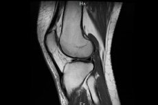
MRI anatomy of the knee joint
It would be useful for patients to know that the knee joint has certain anatomical features. It is a complex mechanism that ensures the connection of the femur with the patella and tibia.
The patella is the anterior joint element that most people know as the "knee cap". It is also important to consider the tendon connections, lateral and cruciate ligaments - this is why the interpretation of the results takes relatively long, but this is the only way to make a correct diagnosis.
The joint cavity contains cruciate ligaments that can be damaged by excessive knee motion. The function of the anterior ligament is to protect the ankle from anterior displacement beyond the permissible limit. This ligament runs through the joint cavity and unites parts of the lower section of the tibia.
On the articular surface there is cartilaginous tissue that forms menisci. The joint system itself is localized in the knee bursa. This mechanism ensures flexion and extension of the limb under different loads.
Most often, patients with ligament ruptures, peritendinous bone fractures, cartilage and meniscus injuries seek MRI assistance. The listed injuries occur with excessive overload of the knee joint, with excessive motor amplitude in different directions.
Such a specific pathology as osteochondritis dissecans of the knee joint on MRI has its own characteristic signs. Most patients have damage to the epiphysis of the femur, specifically the medial condyle. Near the areas of attachment of the posterior cruciate ligament, a defect zone caused by an aseptic necrotic process is determined. The spongy structure in this zone is not traced, the boundaries are usually smooth, relatively clear.
MRI of the knee joint for children
For children of a younger age group, diagnostic MRI is prescribed only if there are compelling indications - as a rule, such a study is carried out using anesthesia.
If a doctor needs to examine the knee joint of an older child, he or she will first talk to the child's parents. It is the parents who should discuss all the details of the examination with the child in advance, and also convince the child that this procedure will not cause any pain or discomfort. If the little patient is afraid of loud sounds, he or she must be warned that it will be noisy while the tomograph is in operation: special headphones will have to be worn.
If the doctor can make a diagnosis without resorting to an MRI of the knee joint, then it is better not to prescribe this type of diagnostics. Most children have difficulty remaining motionless for some time. It is precisely to ensure immobility that small children have to use anesthesia - this is practiced only in extreme, irreplaceable situations.
When evaluating the resulting diagnostic image, the doctor takes into account that the norm for MRI of the knee joint in children has its own characteristics:
- proliferation of blood vessels in the area of the posterior horn of the medial meniscus;
- small volume of fluid in girls;
- subchondrally altered bone tissue.
In children, it is advisable to perform MRI diagnostics of the knee joint on both limbs, even if the child complains of problems on one side.
Contraindications to the procedure
- MRI of the knee joint is not performed on patients with non-removable metal elements in the body, as the latter may be exposed to a magnetic field, heat up and affect the functioning of internal organs. Such elements may include pacemakers, insulin pumps, dental and bone implants, auditory amplifiers, etc.
- The procedure is not entirely suitable as a diagnostic test for people with claustrophobia. Theoretically, diagnostics for such patients is possible in two ways: using an open-type device, and after additional administration of sedatives to the patient.
- Magnetic resonance imaging is not performed on people with mental disorders and a tendency to hyperkinesis. The closed procedure is also not suitable for people suffering from obesity.
- Contrast MRI is not prescribed during pregnancy and breastfeeding, as well as to patients with severe renal insufficiency.
Complications after the procedure
MRI of the knee joint cannot cause any unpleasant consequences for the patient’s health – on the contrary, this examination often allows for a timely and correct diagnosis, which will help not only to maintain the patient’s health, but also to prevent disability.
MRI of the knee never causes complications - on the contrary, this type of examination helps to find hidden diseases that cause many unpleasant symptoms, forcing a person to experience long-term discomfort. It is these diseases, if not detected in time, that can cause serious complications over time, up to and including impaired mobility in the joint and the inability to move normally.
Magnetic resonance imaging is a much safer method than computed tomography or x-rays, that is, diagnostic procedures involving radiation.
Care after the procedure
No special post-diagnostic care for the patient is required after the procedure. After the diagnosis, the person goes home and continues to lead their normal life.
The description of the MRI conclusion of the knee joint is based on the obtained images, which are taken in different projections on different sections. The description is made by the attending physician of the rheumatology, traumatology or orthopedics specialty.
- MRI allows you to describe the condition of the bone tissues that form the joint: bone growths, neoplasms, damage - in particular, cracks, ruptures - are indicated. The use of different sections allows you to track the depth of damage, its size.
- The image perfectly visualizes the structure of cartilage. You can notice symptoms of meniscopathy, changes in the integrity of the cartilage, microscopic damage. Also, the so-called "joint mouse" is determined, which is an element of the meniscus that has detached from it. This condition usually causes a lot of unpleasant symptoms.
- MRI shows in detail the condition of the ligaments, capsular joint fibers. Thanks to this, it is easy to determine the presence of a rupture of the cruciate ligaments, their detachment from the bone. Capsule damage is also diagnosed - for example, the presence of cystic formations, inflammatory processes, etc.
 [ 15 ]
Reviews
Often a person tries to cure a sore joint by resorting to various pills, ointments, compresses, not suspecting that he is treating a completely different disease. To exclude such a situation, you need to do the following: see a doctor and undergo high-quality diagnostics - for example, magnetic resonance imaging. This method will help find a solution to the following knee problems:
- difficulty and discomfort when moving;
- uncomfortable going up and down stairs;
- strange sounds such as crunching or clicking in the knee joint;
- knee pain during or after exercise;
- swelling and inflammation in the knee joint;
- periodic or constant pain in the knee joint without apparent cause.
According to many patients, MRI of the knee joint often helps to find a previously unknown problem, which allows for the treatment that is needed in a particular case.