Medical expert of the article
New publications
Microbiologic and bacterioscopic examination of vaginal discharge
Last reviewed: 07.07.2025

All iLive content is medically reviewed or fact checked to ensure as much factual accuracy as possible.
We have strict sourcing guidelines and only link to reputable media sites, academic research institutions and, whenever possible, medically peer reviewed studies. Note that the numbers in parentheses ([1], [2], etc.) are clickable links to these studies.
If you feel that any of our content is inaccurate, out-of-date, or otherwise questionable, please select it and press Ctrl + Enter.

Microbiological and bacterioscopic examination is used to diagnose inflammatory processes and allows to establish the state of the vaginal biocenosis, as well as some pathogens of sexually transmitted diseases. This examination is carried out during a woman's initial visit to a gynecologist, as well as before gynecological operations and diagnostic manipulations.
To diagnose trichomoniasis, in addition to bacterioscopy of stained smears, vaginal discharge washes are examined with saline solution.
Carrying out bacterioscopy of smears is the leading method in determining the vaginal biocenosis. In healthy women, the biocenosis state is characterized by the predominance of gram-positive lactobacilli (Doderlein bacilli) producing hydrogen peroxide, which creates an acidic environment in the vagina. The acidic reaction of vaginal fluid prevents colonization of the vagina by opportunistic and pathogenic microorganisms. In smears stained according to Gram, a small number of epithelial cells and leukocytes, as well as gram-positive bacilli, are observed.
As a result of changes in the microbial landscape in various diseases, the normocenosis turns into pathological forms: dysbiosis ( bacterial vaginosis ) and vaginitis (colpitis) of various etiologies.
Bacteriological examination
In some cases, when it is necessary to identify microorganisms, secretions from various parts of the reproductive system are sown on appropriate nutrient media. This study is used when there is a suspicion of a specific nature of the inflammatory process and to determine the sensitivity of microflora to antibacterial drugs.
After inserting the vaginal speculums, a metal Volkman spoon is used to scrape the mucous membrane of the cervical canal and the contents are applied to a glass slide in a thin layer as an oblong smear. The speculum is then removed, the urethra is lightly massaged with a finger inserted into the vagina and a scraping of its mucous membrane is made with the other end of the spoon. The scraping is applied to the same glass slide in the form of a round thin smear.
In case of inflammatory processes in the vagina, smears are taken from the posterior fornix with a wooden spatula at the same time as taking smears for flora and applied in a thin, wide layer onto a glass slide.
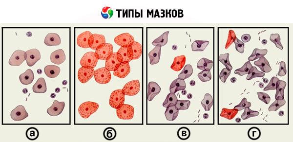
To correctly assess the condition of the vagina, four degrees of purity of vaginal contents are distinguished.
- At the first degree of purity, only Doderlein bacilli and squamous epithelial cells are found in the vaginal smear. The reaction of the contents is acidic.
- The second degree of purity - the smear contains vaginal bacilli, leukocytes (no more than 5 in the field of vision), cocci, epithelium. The reaction is acidic.
- The third degree of purity is characterized by the presence of single Doderlein bacilli in the smear, a large number of various microbes and leukocytes up to 15 in the field of vision. The reaction is neutral.
- The fourth degree - the smear is completely free of Doderlein rods, the entire field of vision is covered with leukocytes, clusters of coccal flora and squamous epithelial cells are detected. The reaction of the contents is alkaline.

For bacteriological examination, the discharge is taken with a sterile cotton swab. To take material from the urethra, the patient should not urinate for 2 hours.
What's bothering you?
What do need to examine?
What tests are needed?


 [
[