Medical expert of the article

New publications
Idiopathic pulmonary hemosiderosis.
Last reviewed: 04.07.2025

All iLive content is medically reviewed or fact checked to ensure as much factual accuracy as possible.
We have strict sourcing guidelines and only link to reputable media sites, academic research institutions and, whenever possible, medically peer reviewed studies. Note that the numbers in parentheses ([1], [2], etc.) are clickable links to these studies.
If you feel that any of our content is inaccurate, out-of-date, or otherwise questionable, please select it and press Ctrl + Enter.
Idiopathic pulmonary hemosiderosis is a lung disease characterized by repeated hemorrhages into the alveoli and a wave-like recurrent course, hypochromic anemia and a wave-like recurrent course.
The etiology and pathogenesis of the disease have not been sufficiently studied. It is assumed that there is a congenital deficiency of the elastic fibers of small and medium pulmonary vessels, which leads to their expansion, blood stasis, and penetration of erythrocytes through the vessel wall. Most researchers consider pulmonary hemosiderosis to be an immunoallergic disease. In response to a sensitizing agent, autoantibodies are formed, an antigen-antibody reaction develops, the shock organ for which is the lungs, which leads to capillary expansion, stasis, and diapedesis of erythrocytes into the lung tissue with the deposition of hemosiderin in it.
Causes idiopathic pulmonary hemosiderosis.
The cause is unknown. It is assumed that there is a congenital deficiency of the elastic fibers of the vessels of the pulmonary circulation, primarily the microcirculatory bed, which leads to dilation of the pulmonary capillaries, a pronounced slowdown in blood flow, diapedesis of erythrocytes into the alveoli, pulmonary parenchyma with subsequent deposition of hemosiderin in it. There is a point of view on the possible role of a congenital anomaly of vascular anastomoses between the bronchial arteries and pulmonary veins.
However, recently the theory of immune complex origin of the disease has become most widespread. It is possible that antibodies are formed to components of the pulmonary vascular wall with subsequent formation of antigen-antibody complexes primarily in the microcirculatory bed of the lungs, which leads to necrosis of the vascular wall with hemorrhages in the alveoli and parenchyma of the lungs. The major role of the cytotoxic effect of immune lymphocytes on the vascular wall is also not excluded.
The following geomorphological changes are characteristic of idiopathic pulmonary hemosiderosis:
- filling of the alveoli with red blood cells;
- detection in the alveoli, alveolar ducts and respiratory bronchioles, as well as in the interstitial tissue of a large number of alveolar macrophages filled with hemosiderin particles;
- thickening of the alveoli and interalveolar septa;
- development of diffuse pneumosclerosis and degenerative changes in the elastic tissue of the lung as the disease progresses;
- disruption of the structure of the basement membrane of the capillaries of the interalveolar septa (according to electron microscopic studies)
Symptoms idiopathic pulmonary hemosiderosis.
Idiopathic pulmonary hemosiderosis may be acute or chronic with repeated exacerbations. Acute course is typical mainly for children.
Complaints of patients during acute or exacerbation of the disease are quite typical. Patients are bothered by cough with separation of bloody sputum. Hemoptysis is one of the main symptoms of the disease and can be expressed significantly (pulmonary hemorrhage). Cases without hemoptysis are very rare. In addition, patients complain of shortness of breath (especially under load), dizziness, tinnitus, flickering flies before the eyes. These complaints are caused mainly by the development of anemia due to prolonged hemoptysis. The development of diffuse pneumosclerosis during the progressive course of the disease is also important in the origin of shortness of breath. Many patients have pain in the chest, joints, abdomen, body temperature rises, significant weight loss is possible.
When remission occurs, the patients' health improves significantly and they may not complain at all or their complaints may be insignificant. The duration of remission varies, but after each exacerbation, as a rule, it decreases.
Where does it hurt?
What's bothering you?
Diagnostics idiopathic pulmonary hemosiderosis.
On examination of patients, attention is drawn to the pallor of the skin and visible mucous membranes, icteric sclera, and cyanosis. The severity of pallor depends on the degree of anemia, and cyanosis - on the degree of respiratory failure. Percussion of the lungs reveals dullness of percussion sound (mainly in the lower parts of the lungs). With extensive hemorrhages in the lung tissue, dullness of percussion sound is much more pronounced and bronchial breathing can be heard above the dull sound zone. Often, such patients, especially in acute or severe exacerbation of the disease, are diagnosed with bilateral pneumonia. Auscultation of the lungs reveals an important sign of idiopathic pulmonary hemosiderosis - widespread crepitation; moist fine-bubble and dry wheezing can be heard. With the development of bronchospastic syndrome, the number of dry wheezing (whistles and buzzing) increases sharply. During auscultation of the heart, attention is drawn to the muffled tones; in the development of chronic pulmonary heart disease, the accent of the second tone on the pulmonary artery is determined; in decompensation of the pulmonary heart disease, the liver enlarges. In 1/3 of patients, liver enlargement is observed even in the absence of decompensated pulmonary heart disease. The spleen may enlarge.
Idiopathic pulmonary hemosiderosis may be complicated by severe infarction pneumonia (it may be extensive and accompanied by severe respiratory failure), recurrent pneumothorax, severe bleeding. These complications may cause death.
Laboratory data
- General blood analysis - hypochromic anemia is typical. It is manifested by a decrease in the hemoglobin level, the number of erythrocytes, the color index, anisocytosis, poikilocytosis. Anemia can be significantly expressed. Reticulocytosis is also observed.
In severe exacerbation of pulmonary hemosiderosis, as well as in the development of infarction pneumonia, severe leukocytosis appears, the leukocyte formula shifts to the left, and ESR increases. Eosinophilia occurs in 10-15% of patients.
- General urine analysis - no significant changes, but sometimes protein and erythrocytes are detected.
- Biochemical blood test - the content of bilirubin, alanine aminotransferase, alpha2- and gamma-globulins increases, the iron content decreases, the total iron-binding capacity of the blood serum increases.
- Immunological studies - no significant changes are detected. In some patients, there may be a decrease in the number of T-lymphocytes, an increase in immunoglobulins, and the appearance of circulating immune complexes.
- Sputum analysis. Erythrocytes and siderophages are detected - alveolar macrophages loaded with hemosiderin. Sputum analysis should be performed frequently, since a single study may be uninformative.
- Study of bronchial lavage fluid - siderophages are found in bronchial lavage waters.
- Bone marrow puncture analysis - myelogram is characterized by a decrease in the number of sideroblasts - red bone marrow cells containing iron lumps. A sign of increased erythropoiesis may be detected - an increase in the number of normoblasts (probably as a manifestation of a compensatory reaction to the development of anemia).
 [ 20 ]
Instrumental research
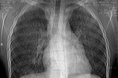
X-ray examination of the lungs. X-ray examination can identify the following stages of the disease:
- Stage I - decreased transparency of the lung tissue (veil-like darkening of both lungs), which is caused by diffuse small hemorrhages into the lung tissue;
- Stage II - manifests itself in the presence of multiple small round foci from 1-2 mm to 1-2 cm in diameter, scattered diffusely throughout all lung fields. These foci gradually resolve within 1-3 weeks. The emergence of new foci coincides with the exacerbation phase of the disease;
- Stage III - characterized by the appearance of extensive intense darkening, which is very reminiscent of infiltrative darkening in pneumonia. The appearance of such intense darkening is due to the development of edema and inflammation around the hemorrhage sites. A characteristic feature of this stage, like the second, is the fairly rapid disappearance and reappearance of infiltrates in other areas of the lungs where the hemorrhage occurred;
- Stage IV - intense interstitial fibrosis is detected, which develops as a result of repeated hemorrhages and the organization of fibrin in the alveoli.
The indicated radiological changes are usually bilateral and are extremely rare when unilateral.
Enlargement of the intrathoracic lymph nodes is uncommon, but may be observed in 10% of patients.
With the development of chronic pulmonary hypertension, a bulging of the pulmonary artery cone and an enlargement of the right sections of the heart are detected. With the development of pneumothorax, a partial or complete collapse of the lung is determined.
Perfusion lung scintigraphy. Idiopathic hemosiderosis is characterized by severe bilateral pulmonary blood flow disturbances.
Study of the ventilation capacity of the lungs. As the disease progresses, restrictive respiratory failure develops, characterized by a decrease in VC. Quite often, a violation of bronchial patency is determined, as evidenced by a decrease in FEV1, the Tiffno index, and peak flowmetry indicators.
ECG. Progressive anemia leads to the development of myocardial dystrophy, which causes a decrease in the amplitude of the T wave in many leads, primarily in the left chest leads. With significantly pronounced myocardial dystrophy, a decrease in the ST interval downwards from the isoline is possible, the appearance of various types of arrhythmia (most often ventricular extrasystole). With the development of chronic pulmonary hypertension, signs of myocardial hypertrophy of the right atrium and right ventricle appear.
Blood gas analysis. With the development of severe respiratory failure, severe arterial hypoxemia appears.
Histological examination of lung tissue biopsy. Lung tissue biopsy (transbronchial, open lung biopsy) is performed in a very limited manner, only when it is absolutely impossible to diagnose the disease. Such a maximum narrowing of indications for lung biopsy is associated with an increased risk of hemorrhage.
Histological examination of lung tissue biopsies reveals a large number of hemosiderophages in the alveoli, as well as pronounced signs of interstitial tissue fibrosis.
Ultrasound examination of abdominal organs. With prolonged existence of the disease, enlargement of the liver and spleen is often detected.
Diagnostic criteria for idiopathic pulmonary hemosiderosis
The main diagnostic criteria for idiopathic pulmonary hemosiderosis can be considered the following:
- repeated and long-term hemoptysis that exists;
- shortness of breath, steadily progressing as the duration of the disease increases;
- fine bubbling diffuse auscultatory manifestations, wheezing;
- a characteristic radiological picture is the sudden appearance of multiple focal shadows throughout all lung fields and their fairly rapid spontaneous disappearance (within 1-3 weeks), the development of interstitial fibrosis;
- detection of siderophages in sputum - alveolar macrophages loaded with hemosiderin;
- hypochromic anemia, decreased iron content in the blood;
- detection of siderophages and interstitial fibrosis in lung tissue biopsies;
- negative tuberculin tests.
Idiopathic Pulmonary Hemosiderosis Screening Program
- General blood and urine tests.
- Biochemical blood test: content of total protein and protein fractions, bilirubin, aminotransferases, seromucoid, fibrin, haptoglobin, iron.
- Immunological studies: content of B- and T-lymphocytes, subpopulations of T-lymphocytes, immunoglobulins, circulating immune complexes.
- Sputum examination: cytological analysis, determination of Mycobacterium tuberculosis, atypical cells, siderophages.
- X-ray examination of the lungs.
- ECG.
- Study of external respiratory function - spirography.
- Ultrasound examination of the heart, liver, spleen, kidneys.
- Study of bronchial lavage fluid: cytological analysis, determination of siderophages.
- Lung biopsy.
Example of formulation of the diagnosis of idiopathic pulmonary hemosiderosis
Idiopathic pulmonary hemosiderosis, acute phase, stage II radiographic, stage II respiratory failure. Chronic iron deficiency anemia of moderate severity.
What do need to examine?
What tests are needed?
Differential diagnosis
Differential diagnostic differences between idiopathic pulmonary hemosiderosis and hematogenous disseminated tuberculosis
Signs |
Idiopathic pulmonary hemosiderosis |
Hematogenous disseminated pulmonary tuberculosis |
Intensity of hemoptysis |
Most often, streaks of blood in the sputum, sometimes intensely blood-stained sputum, severe pulmonary hemorrhage is observed rarely |
Blood streaks in sputum, very often "bloody spitting", "bloody clots", very often - severe pulmonary hemorrhage |
General sputum analysis |
Red blood cells and a large number of siderophages are found - alveolar macrophages filled with hemosiderin |
Many erythrocytes are found, siderophages are not typical and are very rare. |
| Mycobacterium tuberculosis in sputum | Not detected | Are being discovered |
Dynamics of focal lesions in the lungs during X-ray examination |
Spontaneous reverse development is characteristic |
There is no spontaneous reverse development |
The appearance of cavities in the lungs |
Not typical |
Typical |
Lung tissue biopsy examination |
Detection of large numbers of siderophages and interstitial fibrosis |
Siderophages are not detected |
An effective method of treatment |
Glucocorticoid therapy |
Anti-tuberculosis therapy |
Differential diagnosis of idiopathic pulmonary hemosiderosis
- Hematogenous disseminated pulmonary tuberculosis
The main manifestations of hematogenous disseminated pulmonary tuberculosis are described in the article " Pneumonia ". It should be emphasized that there are great differential diagnostic difficulties due to the commonality of the symptoms of the two diseases. Hemoptysis, dyspnea, weakness, weight loss, fine bubbling rales, crepitation, disseminated focal changes in the lungs during X-ray examination are observed both in diopathic hemosiderosis and in hematogenous disseminated pulmonary tuberculosis.
Hemoptysis, anemia, increasing weakness, weight loss force us to differentiate idiopathic pulmonary hemosiderosis from lung cancer. The basic principles of lung cancer diagnostics are described in the article " Pneumonia ". The following signs should also be taken into account:
- in case of cancer, erythrocytes and cancerous (atypical) cells are found in sputum; in case of idiopathic pulmonary hemosiderosis, erythrocytes and siderophages are found;
- in lung cancer, there is never a spontaneous reversal of radiological signs of the disease; in pulmonary hemosiderosis, focal shadows spontaneously disappear with the onset of remission;
- In central lung cancer, expansion and blurring of the contours of the lung root is revealed; for idiopathic hemosiderosis, expansion of the lung roots is not typical.
- Congestive pulmonary hemosiderosis
Pulmonary hemosiderosis may develop as a result of circulatory failure, occurring with congestion in the pulmonary circulation. In this case, hemoptysis may also occur, and crepitation and fine-bubble rales are detected during auscultation of the lungs, and siderophages may be detected in the sputum. Congestive pulmonary hemosiderosis is diagnosed quite simply based on the clinical picture of the underlying heart disease that led to congestion in the lungs (heart defects, dotation cardiomyopathy, cardiosclerosis, etc.) and radiographic signs of congestion in the pulmonary circulation. There is usually no need for a lung biopsy.
- Pneumonia
Hemoptysis, as well as darkening in the lungs in the form of focal infiltration during radiological examination, make it necessary to differentiate idiopathic pulmonary hemosiderosis from pneumonia, including lobar pneumonia.
- Goodpasture's syndrome
The presence of hemoptysis, dyspnea, anemia, and similar auscultatory manifestations make differential diagnostics of pulmonary vdiopathic hemosiderosis and Goodpasture's syndrome necessary. It is presented in the article " Goodpasture's syndrome ".
Treatment idiopathic pulmonary hemosiderosis.
The treatment is carried out as follows.
Glucocorticoid drugs are prescribed. They suppress autoimmune reactions and reduce vascular permeability. Prednisolone is usually used in a daily dose of 30-50 mg. After the condition improves, the dose of prednisolone is gradually reduced (over 3-4 months) to a maintenance dose (5-7.5 mg per day), which is taken for several months.
There is a method of combined treatment with massive plasmapheresis in combination with cytostatics. With the help of plasmapheresis, the produced antibodies are removed from the plasma, and cytostatics reduce the production of new antibodies. Azathioprine and chlorophosphan are usually used. The latter is prescribed at 400 mg every other day, the course of treatment is 8-10 g.
Combined treatment with prednisolone, iron preparations in combination with anticoagulant and antiplatelet agents (heparin, curantil, trental) is effective.
Due to the development of iron deficiency anemia, patients should regularly take iron-containing drugs - ferroplex, tardiferon, conferon, etc.
In the development of chronic pulmonary heart disease, treatment is carried out aimed at reducing pulmonary hypertension.