Medical expert of the article
New publications
Bronchial and tracheal examination
Last reviewed: 07.07.2025

All iLive content is medically reviewed or fact checked to ensure as much factual accuracy as possible.
We have strict sourcing guidelines and only link to reputable media sites, academic research institutions and, whenever possible, medically peer reviewed studies. Note that the numbers in parentheses ([1], [2], etc.) are clickable links to these studies.
If you feel that any of our content is inaccurate, out-of-date, or otherwise questionable, please select it and press Ctrl + Enter.

The trachea and bronchi belong to the lower respiratory tract and provide the function of external respiration, therefore the main symptom of their various pathological conditions is often the insufficiency of external respiration, developing as a result of obstruction of the airways.
When examining a patient with a respiratory disease, the doctor must first assess the state of external respiration, for which he pays attention to the patient's behavior and appearance, identifies signs of hypoxia, and only then proceeds to the anamnesis and special instrumental research methods.
The behavior of a patient with a lesion of the lower respiratory tract in some cases allows us to judge the nature of the disease or, at least, to determine the direction of the diagnostic search. In case of stenosis of the respiratory tract, as well as in other disorders of the external respiratory function ( bronchial asthma, pulmonary edema, atelectasis ), the patient, as a rule, takes a forced sitting position with support on the arms and a slightly forward-leaning body. The patient also takes this position in case of respiratory failure due to paralysis of the respiratory muscles (various myoplegic syndromes).
The appearance of the patient's face has a certain significance for assessing the patient's condition. For example, in former times, descriptions included such a concept as the "Venetian face", characteristic of patients suffering from pulmonary tuberculosis for a long time.
Such patients are characterized by transparent pallor of the skin, sunken eyes with a feverish shine and circles of blue, a deep sad look of a doomed person. "Restless face" - an open mouth, an anxious wandering look, the head is raised, the neck is stretched out. This appearance is typical for patients suffering from an attack of bronchial asthma, left ventricular heart failure or severe bronchopneumonia. "Cyanotic face" - cyanosis of the lips, nose, cheeks, pale-cyanotic spots on the sides of the wings of the nose; these signs can have many causes: severe bronchopneumonia with obstruction of the bronchi and bronchioles, circulatory failure, cardiopulmonary failure. Facial cyanosis also appears with tumors or diverticula of the esophagus, compressing the lower respiratory tract, with incomplete obstruction of the trachea or one of the main bronchi by a foreign body, with exudative pleurisy or severe ascites, limiting respiratory excursions of the lungs, etc.
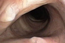
Local examination of the trachea and bronchi includes endoscopy and radiography. The first is performed using special optical devices - bronchoscopes, the second - by the generally accepted method of X-ray diagnostics.
Other methods of examining the tracheobronchial system include radiological, cytological, biopsy and gas mediastinography.
How to examine?


 [
[