Medical expert of the article
New publications
Ichthyoses
Last reviewed: 05.07.2025

All iLive content is medically reviewed or fact checked to ensure as much factual accuracy as possible.
We have strict sourcing guidelines and only link to reputable media sites, academic research institutions and, whenever possible, medically peer reviewed studies. Note that the numbers in parentheses ([1], [2], etc.) are clickable links to these studies.
If you feel that any of our content is inaccurate, out-of-date, or otherwise questionable, please select it and press Ctrl + Enter.

Ichthyosis is a group of hereditary skin diseases characterized by impaired keratinization.
 [ 1 ]
[ 1 ]
Causes and pathogenesis of ichthyosis
The causes and pathogenesis are not fully understood. Many forms of ichthyosis are based on mutations or disturbances in the expression of genes encoding various forms of keratin. In lamellar ichthyosis, there is a deficiency of keratinocyte transglutaminase and proliferative hyperkeratosis. In X-linked ichthyosis, there is a deficiency of sterol sulfatase.
Pathomorphology of ichthyosis
Characterized by hyperkeratosis, thinning or absence of granular, thinning of spinous layers of the epidermis. Hyperkeratosis often extends to the mouths of hair follicles, which is clinically manifested by follicular keratosis.
The spinous layer shows signs of slight atrophy and consists of small, atrophic epithelial cells or, on the contrary, large cells with vacuolization phenomena. The amount of melanin in the basal layer is sometimes increased. Mitotic activity is normal or decreased. The number of hair follicles is reduced, the sebaceous glands are atrophic.
In the dermis, the number of small vessels is increased; small perivascular infiltrates consisting of lymphoid cells and tissue basophils can be detected.
Histogenesis of ichthyosis
Thinning or absence of the granular layer is caused by a defect in the synthesis of keratohyalin, the granules of which appear small, fine-grained or spongy in electron microscopic images, localized at the edges of the tonofilament bundles. In the stratum corneum the cells are flattened, dissolution of their desmosomes occurs in the 25-35th row (normally this process occurs in the 4th-8th row), which is morphologically manifested by a delay in the rejection of stratum corneum cells and an increase in the adhesiveness of stratum corneum cells. The basis of the violation of the keratinization process is a defect in the synthesis of the main protein of the epidermis - keratohyalin, a violation of its normal polymerization, which can be the result of an incorrect sequence of amino acids in the polypeptide chain, the loss of one of its components or a change in the amount, as well as a violation of the activity of specific keratinization enzymes. However, the relationship between the defect in keratohyalin synthesis and a change in the adhesive properties of horny scales is still unclear. The presence of a combined genetic defect cannot be ruled out.
Symptoms of Ichthyosis
The following forms of ichthyosis are distinguished: common, X-linked, lamellar and epidermolytic.
Ichthyosis vulgaris is the most common form of the disease and is inherited in an autosomal dominant manner.
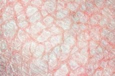
Clinically, it usually manifests itself by the end of the first year of life as dry skin, follicular keratosis, flaking with the presence of light, tightly attached polygonal scales resembling "fish scales". There are no inflammatory phenomena. The extensor surfaces of the extremities, the back, and to a lesser extent the abdomen are affected mainly, there are no changes in the folds of the skin.
The skin of the palms and soles looks senile due to the increased papillary pattern and deepening of the skin folds.
Common ichthyosis begins in early childhood (3-12 months) and is inherited in an autosomal dominant manner. Men and women are equally affected. The skin pathological process is characterized by dryness and flaking of the skin, most pronounced on the extensor surfaces of the extremities, follicular hyperkeratosis. On the palms and soles, the skin pattern is enhanced, occasionally - keratoderma. The skin is pale, with a yellowish-gray tint, covered with abundant dry or larger, polygonal, grayish-white or more often dirty-gray, translucent in the center and peeling off at the edges scales, which will give the skin a cracked, bran-like appearance. Some patients have large-plate peeling in the form of fish scales. The scalp is dry, abundantly covered with bran-like scales (as if sprinkled with flour). The hair is dry, thinned, dull. The nails of most patients are not changed, but sometimes dystrophic changes are observed. Subjective disorders are usually absent, but there may be itching with pronounced dryness, more often in winter, when an exacerbation of the disease is often noted. The frequency of atopy is increased. The general health of most patients is not impaired. The disease, although somewhat relieved with age, exists throughout life, weakening in the summer.
What's bothering you?
X-linked recessive ichthyosis
X-linked recessive ichthyosis (synonym: black ichthyosis, ichthyosis nigricans). occurs with a frequency of 1:6000 in males, the type of inheritance is recessive, sex-linked, the full clinical picture is observed only in males. It can exist from birth, but most often appears in the first weeks or months of life. The skin is covered with large, brownish, tightly attached thick scales, localized mainly on the anterior surface of the trunk, head, neck, flexor and extensor surfaces of the limbs. Skin lesions are often accompanied by corneal opacity, hypogonadism, cryptorchidism. Unlike normal ichthyosis, an earlier onset of the disease is noted, there are no changes in the palms and soles, skin folds are affected, manifestations of the disease are more pronounced on the flexor surfaces of the limbs and on the abdomen. As a rule, follicular keratosis is absent.
X-linked recessive ichthyosis is less common than usual, and the manifestations of the disease may be similar to those observed in usual ichthyosis. However, this form of ichthyosis is characterized by a number of distinctive clinical signs. Dermatosis begins in the first weeks or months of life, but may exist from birth. The skin is dry, covered with finely plated, sometimes larger and thick, tightly adhering to the surface, dark brown to black scales. The process is localized on the trunk, especially on the extensor surfaces of the limbs. The palms and soles are not affected, follicular hyperkeratosis is absent. Clouding of the cornea is noted in 50% of patients and cryptorchidism in 20%.
Pathomorphology. The main histological sign is hyperkeratosis with a normal or slightly thickened granular layer. The stratum corneum is massive, reticular, in places many times thicker than normal. The granular layer is represented by 2-4 rows of cells, with electron microscopic examination revealing keratohyaline granules of normal size and shape. The number of lamellar granules is reduced. The melanin content in the basal layer is increased. The proliferative activity of the epidermis is not impaired, the transit time is slightly increased compared to the norm. As a rule, no changes are detected in the dermis.
Histogenesis. As in the usual form of the disease, in X-linked ichthyosis, retention hyperkeratosis is broken down, but its origin is different. The main genetic defect in this form of ichthyosis is a deficiency of sterol sulfatase (steroid sulfatase), the gene of which is located in the Xp22.3 locus. Steroid sulfatase hydrolyzes sulfur esters of 3-beta-hydroxysteroids, including cholesterol sulfate and a number of steroid hormones. In the epidermis, cholesterol sulfate, produced from cholesterol, is located in the intercellular spaces of the granular layer. Due to disulfide bridges and lipid polarization, it participates in the stabilization of membranes. Its hydrolysis promotes exfoliation of the stratum corneum, since glycosidase and sterol sulfatase contained in this layer participate in intercellular adhesion and desquamation. It is obvious that in the absence of sterol sulfatase, intercellular connections are not weakened and retention hyperkeratosis develops. In this case, a high content of cholesterol sulfate is found in the stratum corneum. A decrease or absence of sterol sulfatase was found in the culture of fibroblasts and epithelial cells, hair follicles, neurofilaments and leukocytes of patients. An indirect indicator of their deficiency can be the acceleration of electrophoresis of 3-lipoproteins of blood plasma. Enzyme deficiency is also determined in women - gene carriers. Antenatal diagnostics of this type of ichthyosis is possible by determining the content of estrogens in the urine of pregnant women. Aryl sulfatase C of the placenta hydrolyzes dehydroepiandrosterone sulfate, produced by the adrenal glands of the fetus, which is a precursor of estrogens. In the absence of the above-mentioned enzyme, the content of estrogens in the urine decreases. Observation of the development of children born to mothers with hormone deficiency showed that ethnic manifestations of X-linked ichthyosis are variable. With the help of biochemical study of enzymes, X-linked ichthyosis was diagnosed in some cases in patients with the clinical picture of ordinary ichthyosis. X-linked ichthyosis can be part of more complex genetically determined syndromes. A case of a combination of skin lesions typical of X-linked ichthyosis with short stature and mental retardation associated with translocation of the Xp22.3-pter segment of the chromosome is described.
Syndromes that include ichthyosis as one of the symptoms include, in particular, Refsum and Podlit syndromes.
Refsum syndrome
Refsum syndrome includes, in addition to skin changes resembling common ichthyosis, cerebellar ataxia, peripheral neuropathy, retinitis pigmentosa, sometimes deafness, eye and skeletal changes. Histological examination reveals, along with signs of common ichthyosis, vacuolization of the cells of the basal layer of the epidermis, in which fat is detected when stained with Sudan III.
The basis of histogenesis is a defect expressed in the inability to oxidize phytanic acid. Normally, it is not detected in the epidermis, but in Refsum syndrome it accumulates, constituting a significant part of the lipid fraction of the intercellular substance, which leads to a violation of the adhesion of horny scales and their exfoliation and a violation of the formation of arachidonic acid metabolites, in particular prostaglandins, involved in the regulation of epidermal proliferation, resulting in the development of proliferative gynerkeratosis and acanthosis.
Podlit syndrome
Podlit syndrome, in addition to skin changes of the type of common ichthyosis, includes hair anomalies (twisted hair, nodular trichorrhexis) with their sparseness, dystrophic changes in the nail plates, dental caries, cataracts, mental and physical retardation. It is inherited in an autosomal recessive manner, the basis of the development of the process is a defect in the synthesis, transport or assimilation of sulfur-containing amino acids. Histological changes are the same as in common ichthyosis.
Treatment. General treatment consists of neotigazone at a daily dose of 0.5-1.0 mg/kg or vitamin A in high doses, emollients and keratolytics are applied locally.
Lamellar ichthyosis
Lamellar ichthyosis is a rare, severe disease, inherited in most cases in an autosomal recessive manner. Some patients have a defect in epidermal transglutaminase. Clinical manifestations at birth include "collodion fetus" or diffuse erythema with lamellar scaling.
Lamellar (plate) ichthyosis
Lamellar (plate) ichthyosis exists from birth and is severe. Children are born in a horny "shell" (collodion fetus) of large, thick, dark-colored plate-like scales separated by deep cracks. The skin-pathological process is widespread and affects the entire skin, including the face, scalp, palms and soles. Most patients have pronounced ectropion and ear deformation. The palms and soles have massive keratosis with cracks, limiting the movement of small joints. Dystrophy of the nails and nail plates is noted, often according to the opichogryphosis type. Sweat and sebum secretion are reduced. There is pronounced peeling on the scalp, the hair is stuck together with scales, and its thinning is noted. Cicatricial alopecia is observed due to the addition of a secondary infection. Dermatosis can be combined with various developmental anomalies (short stature, deafness, blindness, etc.). The disease lasts throughout life.
The disease is transmitted in an autosomal dominant manner, can be congenital or begin soon after birth. Men and women are equally affected. Soon after birth, blisters appear, opening up, they form erosions, which in turn heal without leaving traces. Then keratinization of the skin develops up to wart-like layers in the skin folds, elbows and popliteal fossae. The scales are dark, tightly attached to the skin and usually form a pattern similar to velvet. The rash is accompanied by an odor. The repeated appearance of blisters on the keratinized skin, as well as the exfoliation of horny layers lead to the fact that the skin acquires an absolutely normal appearance. There are also islands of normal skin in the middle of the keratinization foci - this is a characteristic diagnostic sign. The process is localized only on the skin of the folds, palms and soles. The hair is not changed, deformation of the nails is possible.
Collodion fruit
Collodion fetus (syn.: ichthyosis sebacea, seborrhea squamosa neonatorum) is an expression of various disorders of the keratinization process. In most cases (60%), collodion fetus precedes non-bullous recessive ichthyosiform erythroderma. At birth, the baby's skin is covered with a film of tightly adjoining inelastic scales resembling collodion. Under the film, the skin is red, in the fold area there are cracks from which peeling begins, continuing from the first day of life to 18-60 days. Ectropion, exlabion, changes in the shape of the auricles are often observed, the fingers are fixed in a semi-bent position with the thumb extended. In 9.7% of cases, the condition of collodion fetus resolves without any consequences.
Patients with normal-appearing skin at birth have been described, but in such cases X-linked ichthyosis must be excluded. Usually the entire body, including skin folds, is covered with large, yellowish, sometimes dark, saucer-shaped scales against the background of erythroderma. Almost all patients have pronounced ectropion, diffuse keratoderma of the palms and soles, increased hair and nail growth with deformation of the nail plates. Less common are baldness, brachy- and syndactyly of the feet, short stature, deformation and small size of the auricles, cataracts.
Pathomorphology. Moderate acanthosis, papillomatosis (simultaneous proliferation of dermal and epidermal papillae), widening of epidermal outgrowths and pronounced hyperkeratosis are detected in the epidermis. The thickness of the stratum corneum is 2 times greater than the thickness of the entire epidermis under normal conditions; focal parakeratosis is observed in rare cases. The granular layer is mostly unchanged, although it is sometimes thickened. The spinous and basal layers have increased mitotic activity associated with increased proliferation of epithelial cells, the transit time of which is shortened to 4-5 days. Electron microscopy reveals increased metabolic activity of epithelial cells, as evidenced by an increase in the number of mitochondria and ribosomes in their cytoplasm. Electron-transparent crystals located intracellularly are detected in the stratum corneum, and electron-dense clusters are found along the cytoplasmic membranes. In some places there are areas of incomplete keratinization with the presence of remains of destroyed organelles and lipid inclusions. Between the horny scales and the granular layer there are 1-2 rows of parakeratotic cells. Keratohyalin granules are contained in approximately 7 rows of cells, in the intercellular spaces there are numerous lamellar granules.
Histogenesis. The pathological process is based on the inability of epithelial cells to form a marginal band in the stratum corneum, i.e. the outer shell of squamous epithelial cells. Electron-transparent crystals, according to L. Kanerva et al. (1983), are cholesterol crystals. Along with autosomal recessive lamellar ichthyosis, an autosomal dominant variant has been described, which is similar in clinical and histological features. However, its distinctive feature is the presence of a wider layer of parakeratotic cells, which indicates a slowdown in the keratinization process. The structure of the stratum corneum is unchanged.
Treatment: Same as for X-linked ichthyosis.
 [ 4 ], [ 5 ], [ 6 ], [ 7 ], [ 8 ], [ 9 ], [ 10 ]
Epidermolytic ichthyosis
Epidermolytic ichthyosis (synonyms: congenital bullous ichthyosiform erythroderma of Brocq, bullous ichthyosis, etc.)
Histopathology: The granular layer shows giant keratohyalin granules and vacuolization, cell lysis and formation of subcorneal multilocular bullae, as well as papillomatosis and hyperkeratosis.
Treatment: The same as for other forms of ichthyosis.
What do need to examine?
How to examine?
Who to contact?
Treatment of ichthyosis
Emollients and keratolytic agents are prescribed.

