Medical expert of the article

New publications
Examination of subcutaneous adipose tissue
Last reviewed: 05.07.2025

All iLive content is medically reviewed or fact checked to ensure as much factual accuracy as possible.
We have strict sourcing guidelines and only link to reputable media sites, academic research institutions and, whenever possible, medically peer reviewed studies. Note that the numbers in parentheses ([1], [2], etc.) are clickable links to these studies.
If you feel that any of our content is inaccurate, out-of-date, or otherwise questionable, please select it and press Ctrl + Enter.

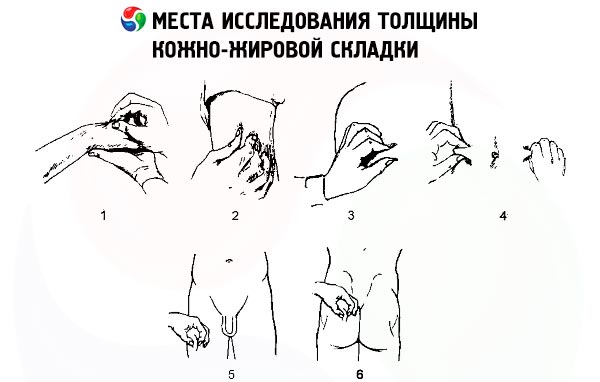
The subcutaneous fat layer is examined almost simultaneously with the skin. The degree of development of the fatty tissue is often in accordance with the body weight and is determined by the size of the skin fold on the abdomen in the navel area; with a sharp decrease in it, the skin is easier to take into a fold, with significant fat deposits this is often impossible to do.
The detection of edema is of great clinical importance.
Edema
Edema (fluid retention) occurs primarily in the subcutaneous tissue due to its porous structure, especially where the tissue is looser. Hydrostatic and hydrodynamic factors explain the occurrence of edema in low-lying areas of the body (lower limbs). The latter factor plays an important role in the development of edema in heart diseases accompanied by congestive heart failure. Edema occurs more often by the end of the day, when the patient has been in an upright position for a long time. At the same time, in kidney diseases, minor edema often appears primarily on the face (in the eyelid area) and usually in the morning. In this regard, the patient may be asked whether he feels heaviness or swelling of the eyelids in the morning. Relatives of the patient may be the first to notice the appearance of such edema.

In diseases of the heart, kidneys, liver, intestines, endocrine glands, edemas can be widespread. In case of venous and lymphatic outflow disorders, allergic reactions, edemas are often asymmetrical. In rare cases, in elderly people, they can appear during prolonged stay in a vertical position, which (like edemas in women in hot weather) is not of great clinical significance.
Patients may seek medical attention with complaints of joint swelling, facial and leg edema, rapid weight gain, and shortness of breath. With general fluid retention, edema occurs primarily, as already mentioned, in low-lying parts of the body: in the lumbosacral region, which is especially noticeable in people in an upright or semi-recumbent position. This situation is typical of congestive heart failure. If the patient can lie in bed, edema occurs primarily on the face and arms, as is the case with young people with kidney disease. Fluid retention is caused by increased venous pressure in any area, for example, in pulmonary edema due to left ventricular failure or in the development of ascites in patients with increased pressure in the portal vein system ( portal hypertension ).
Usually, the development of edema is accompanied by an increase in body weight, but even initial edema in the legs and lower back is easily detected by palpation. It is most convenient to press the tissue to the dense surface of the tibia with two or three fingers, and after 2-3 seconds, if edema is present, pits are detected in the subcutaneous fat tissue. A weak degree of edema is sometimes referred to as "pastosity". Pits on the shin are formed by pressure only if body weight has increased by at least 10-15%. In chronic lymphoid edema, myxedema (hypothyroidism), edema is more dense, and a pit is not formed by pressure.
In both general and local edema, factors involved in the formation of interstitial fluid at the capillary level play an important role in their development. Interstitial fluid is formed as a result of its filtration through the capillary wall - a kind of semipermeable membrane. Some of it returns back to the vascular bed due to the drainage of the interstitial space through the lymphatic vessels. In addition to the hydrostatic pressure inside the vessels, the filtration rate of fluid is affected by the osmotic pressure of proteins in the interstitial fluid, which is important in the formation of inflammatory, allergic and lymphatic edema. Hydrostatic pressure in the capillaries varies in different parts of the body. Thus, the average pressure in the pulmonary capillaries is about 10 mm Hg, while in the renal capillaries it is about 75 mm Hg. When the body is in an upright position, due to gravity, the pressure in the capillaries of the legs is higher than in the capillaries of the head, which creates conditions for the appearance of mild edema of the legs by the end of the day in some people. The pressure in the capillaries of the legs of a person of average height in a standing position reaches 110 mm Hg.
Severe general edema (anasarca) can occur with hypoproteinemia, in which oncotic pressure, mainly associated with the albumin content in plasma, falls, and fluid is retained in the interstitial tissue without entering the vascular bed (often a decrease in the amount of circulating blood is observed - oligemia, or hypovolemia).
The causes of hypoproteinemia can be a variety of conditions, clinically united by the development of edema syndrome. These include the following:
- insufficient protein intake (starvation, poor nutrition);
- digestive disorders (impaired secretion of enzymes by the pancreas, for example, in chronic pancreatitis, other digestive enzymes);
- impaired absorption of food products, primarily proteins (resection of a significant portion of the small intestine, damage to the wall of the small intestine, gluten enteropathy, etc.);
- impaired albumin synthesis (liver disease);
- significant loss of protein in the urine in nephrotic syndrome;
- loss of protein through the intestines (exudative enteropathy ).
The decrease in intravascular blood volume associated with hyperproteinemia may cause secondary hyperaldosteronism via the renin-angiotensin system, which promotes sodium retention and edema formation.
Heart failure causes swelling due to the following reasons:
- disturbance of venous pressure, which can be detected by dilated veins in the neck;
- hyperaldosteronism effect;
- renal blood flow disorder;
- increased secretion of antidiuretic hormone;
- decrease in oncotic pressure due to blood stagnation in the liver, decreased albumin synthesis, decreased protein intake due to anorexia, loss of protein in the urine.
Renal edema is most pronounced in nephrotic syndrome, when, due to pronounced proteinuria, a significant amount of protein is lost (primarily albumin), which leads to hypoproteinemia and hypooncotic fluid retention. The latter is aggravated by developing hyperaldosteronism with increased renal reabsorption of sodium. The mechanism of edema development in acute nephritic syndrome is more complex (for example, at the height of typical acute glomerulonephritis ), when, apparently, a more significant role is played by the vascular factor (increased permeability of the vascular wall), in addition, sodium retention is important, leading to an increase in the volume of circulating blood, "blood edema" (hypervolemia, or plethora). As in heart failure, edema is accompanied by a decrease in diuresis (oliguria) and an increase in the patient's body weight.
Local edema may be caused by venous, lymphatic or allergic factors, as well as local inflammatory processes. With external compression of veins, venous thrombosis, venous valve insufficiency, varicose veins, capillary pressure in the corresponding area increases, which leads to blood stagnation and edema. Most often, thrombosis of the veins of the legs develops in diseases that require prolonged bed rest, including conditions after surgery, as well as during pregnancy.
When lymph drainage is delayed, water and electrolytes are reabsorbed back into the capillaries from the interstitial tissue, but proteins filtered from the capillary into the interstitial fluid remain in the interstitium, which is accompanied by water retention. Lymphatic edema also occurs as a result of obstruction of the lymphatic pathways by filariae ( filariasis is a tropical disease). Both legs and external genitalia can be affected. The skin in the affected area becomes rough, thickened, and elephantiasis develops.
In a local inflammatory process, as a result of tissue damage (infection, ischemia, exposure to certain chemicals such as uric acid), histamine, bradykinin and other factors are released, which cause vasodilation and increased capillary permeability. The inflammatory exudate contains a large amount of protein, which disrupts the mechanism of tissue fluid movement. Often, classic signs of inflammation are observed simultaneously, such as redness, pain, and local increase in temperature.
Increased capillary permeability is also observed in allergic conditions, but unlike inflammation there is no pain and no redness. In Quincke's edema - a special form of allergic edema (usually on the face and lips) - symptoms usually develop so quickly that life is threatened due to swelling of the tongue, larynx, neck (asphyxia).
Disruption of subcutaneous fat tissue development
When examining subcutaneous fat tissue, attention is usually drawn to its increased development. In obesity, excess fat is deposited in the subcutaneous tissue fairly evenly, but to a greater extent in the abdominal area. Uneven deposition of excess fat is also possible. The most typical example is Cushing's syndrome (observed with excessive secretion of corticosteroid hormones by the adrenal cortex), Cushingoid syndrome is often noted, associated with long-term treatment with corticosteroid hormones. Excess fat in these cases is deposited mainly on the neck, face, and upper body, the face usually looks rounded, and the neck is full (the so-called moon face).
The skin of the abdomen often stretches significantly, which is manifested by the formation of areas of atrophy and scars of a purple-blue color, in contrast to the whitish areas of skin atrophy from stretching after pregnancy or large edemas.
Progressive lipodystrophy and significant loss of subcutaneous fat (as well as mesenteric fat) are possible, which is observed in a number of serious diseases, after major surgical interventions, especially on the gastrointestinal tract, during starvation. Local atrophy of subcutaneous fat is observed in patients with diabetes mellitus at the sites of insulin injection. Often, muscle mass of the body decreases simultaneously. The extreme degree of such weight loss is called cachexia.
 [ 5 ], [ 6 ], [ 7 ], [ 8 ], [ 9 ], [ 10 ], [ 11 ], [ 12 ], [ 13 ], [ 14 ]