Medical expert of the article

New publications
Primary hyperaldosteronism - Overview of information
Last reviewed: 12.07.2025

All iLive content is medically reviewed or fact checked to ensure as much factual accuracy as possible.
We have strict sourcing guidelines and only link to reputable media sites, academic research institutions and, whenever possible, medically peer reviewed studies. Note that the numbers in parentheses ([1], [2], etc.) are clickable links to these studies.
If you feel that any of our content is inaccurate, out-of-date, or otherwise questionable, please select it and press Ctrl + Enter.
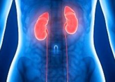
Primary aldosteronism (Conn's syndrome) is aldosteronism caused by autonomous production of aldosterone by the adrenal cortex (due to hyperplasia, adenoma, or carcinoma). Symptoms and signs include episodic weakness, increased blood pressure, and hypokalemia. Diagnosis includes determination of plasma aldosterone levels and plasma renin activity. Treatment depends on the cause. The tumor is removed if possible; in hyperplasia, spironolactone or related drugs may normalize blood pressure and cause the disappearance of other clinical manifestations.
Aldosterone is the most potent mineralocorticoid produced by the adrenal glands. It regulates sodium retention and potassium loss. In the kidneys, aldosterone causes sodium to be transferred from the lumen of the distal tubules to the tubular cells in exchange for potassium and hydrogen. The same effect is observed in the salivary, sweat glands, intestinal mucosal cells, and the exchange between intracellular and extracellular fluid.
Aldosterone secretion is regulated by the renin-angiotensin system and, to a lesser extent, by ACTH. Renin, a proteolytic enzyme, accumulates in the juxtaglomerular cells of the kidneys. A decrease in the volume and velocity of blood flow in the afferent renal arterioles induces renin secretion. Renin converts hepatic angiotensinogen to angiotensin I, which is transformed by angiotensin-converting enzyme into angiotensin II. Angiotensin II causes the secretion of aldosterone and, to a lesser extent, the secretion of cortisol and deoxycorticosterone, which also have pressor activity. Sodium and water retention caused by increased aldosterone secretion increase circulating blood volume and decrease renin secretion.
The syndrome of primary hyperaldosteronism was described by J. Conn (1955) in connection with an aldosterone-producing adenoma of the adrenal cortex (aldosteroma), the removal of which led to a complete recovery of the patient. At present, the collective concept of primary hyperaldosteronism unites a number of diseases that are similar in clinical and biochemical signs, but different in pathogenesis, which are based on excessive and independent (or partially dependent) on the renin-angiotensin system production of aldosterone by the adrenal cortex.
 [
[ What causes primary aldosteronism?
Primary aldosteronism may be caused by an adenoma, usually unilateral, of the cells of the glomerular layer of the adrenal cortex or, less commonly, by adrenal carcinoma or hyperplasia. In adrenal hyperplasia, which is more common in older men, both adrenals are hyperactive and there is no adenoma. The clinical picture may also be seen in congenital adrenal hyperplasia due to 11-hydroxylase deficiency and in dominantly inherited dexamethasone-suppressible hyperaldosteronism.
Symptoms of Primary Aldosteronism
Hypernatremia, hypervolemia, and hypokalemic alkalosis may occur, causing episodic weakness, paresthesias, transient paralysis, and tetany. Diastolic hypertension and hypokalemic nephropathy with polyuria and polydipsia are common. In many cases, mild to moderate hypertension is the only manifestation. Edema is uncommon.
Symptoms of Primary Hyperaldosteronism
Clinical case of primary hyperaldosteronism
Patient M., a 43-year-old woman, was admitted to the endocrinology department of the Kazan Republican Clinical Hospital on January 31, 2012 with complaints of headaches, dizziness with an increase in blood pressure, up to a maximum of 200/100 mm Hg (with a comfortable blood pressure of 150/90 mm Hg), generalized muscle weakness, leg cramps, general weakness, and rapid fatigue.
History of the disease. The disease developed gradually. For five years, the patient has noted increased blood pressure, for which she was observed by a therapist at her place of residence, received antihypertensive therapy (enalapril). About 3 years ago, she began to experience periodic pain in the legs, cramps, muscle weakness, occurring without visible provoking factors, passing on their own within 2-3 weeks. Since 2009, she has received inpatient treatment 6 times in the neurological departments of various healthcare institutions with the diagnosis: Chronic demyelinating polyneuropathy, subacute developing generalized muscle weakness. One of the episodes was with weakness of the neck muscles and drooping of the head.
With the infusion of prednisolone and polarizing mixture, improvement occurred within a few days. According to blood tests, potassium is 2.15 mmol/l.
From 26.12.11 to 25.01.12 she was undergoing inpatient treatment at the Republican Clinical Hospital, where she was admitted with complaints of generalized muscle weakness and periodic leg cramps. An examination was performed, which revealed the following: blood test on 27.12.11: ALT - 29 U/l, AST - 14 U/l, creatinine - 53 μmol/l, potassium 2.8 mmol/l, urea - 4.3 mmol/l, total. Protein 60 g/l, total bilirubin - 14.7 μmol/l, CPK - 44.5, LDH - 194, phosphorus 1.27 mmol/l, Calcium - 2.28 mmol/l.
Urine analysis from 12/27/11; specific gravity - 1002, protein - traces, leukocytes - 9-10 in the field of view, epithelial cells - 20-22 in the field of view.
Hormones in the blood: free T3 - 4.8, free T4 - 13.8, TSH - 1.1 μE/l, cortisol - 362.2 (normal 230-750 nmol/l).
Ultrasound: Kidneys left: 97x46 mm, parenchyma 15 mm, increased echogenicity, renal pelvic floor surface area - 20 mm. Increased echogenicity. The cavity is not dilated. Right 98x40 mm. Parenchyma 16 mm, increased echogenicity, renal pelvic floor surface area 17 mm. Increased echogenicity. The cavity is not dilated. A hyperechoic rim is visualized around the pyramids on both sides. Based on the physical examination and laboratory test data, further examination was recommended to exclude endocrine pathology of adrenal genesis.
Ultrasound of the adrenal glands: in the projection of the left adrenal gland, an isoechoic round formation of 23x19 mm is visualized. In the projection of the right adrenal gland, pathological formations are not reliably visualized.
Urine for catecholamines: Diuresis - 2.2 l, adrenaline - 43.1 nmol/day (normal 30-80 nmol/day), noradrenaline - 127.6 nmol/l (normal 20-240 nmol/day). These results excluded the presence of pheochromocytoma as a possible cause of uncontrolled hypertension. Renin from 13.01.12 - 1.2 μIU/ml (N vert- 4.4-46.1;, horiz 2.8-39.9), aldosterone 1102 pg/ml (normal: lying 8-172, sitting 30-355).
CT scan from 18.01.12: CT signs of a formation in the left adrenal gland (an isodense formation of an oval shape measuring 25*22*18 mm, homogeneous, with a density of 47 HU is determined in the medial peduncle of the left adrenal gland.
Based on the anamnesis, clinical picture, laboratory and instrumental research data, the clinical diagnosis was established: Primary hyperaldosteronism (aldosteronoma of the left adrenal gland), first detected as hypokalemic syndrome, neurological symptoms, sinus tachycardia. Hypokalemic periodic convulsions with generalized muscle weakness. Hypertension grade 3, stage 1. CHF 0. Sinus tachycardia. Urinary tract infection in the resolution stage.
Hyperaldosteronism syndrome occurs with clinical manifestations caused by three main symptom complexes: arterial hypertension, which can have both a crisis course (up to 50%) and persistent; impaired neuromuscular conduction and excitability, which is associated with hypokalemia (in 35-75% of cases); impaired renal tubular function (50-70% of cases).
The patient was recommended surgical treatment to remove the hormone-producing tumor of the adrenal gland - laparoscopic left adrenalectomy. The operation was performed - laparoscopic left adrenalectomy in the abdominal surgery department of the Republican Clinical Hospital. The postoperative period was uneventful. On the 4th day after the operation (11.02.12), the blood potassium level was 4.5 mmol/l. BP 130/80 mm Hg.
Secondary aldosteronism
Secondary aldosteronism is increased adrenal production of aldosterone in response to nonpituitary, extraadrenal stimuli, including renal artery stenosis and hypovolemia. Symptoms are similar to those of primary aldosteronism. Treatment involves correction of the underlying cause.
Secondary aldosteronism is caused by decreased renal blood flow, which stimulates the renin-angiotensin mechanism with resultant hypersecretion of aldosterone. Causes of decreased renal blood flow include obstructive diseases of the renal artery (eg, atheroma, stenosis), renal vasoconstriction (in malignant hypertension), and diseases associated with edema (eg, heart failure, cirrhosis with ascites, nephrotic syndrome). Secretion may be normal in heart failure, but hepatic blood flow and aldosterone metabolism are decreased, so circulating hormone levels are high.
Diagnosis of primary aldosteronism
The diagnosis is suspected in patients with hypertension and hypokalemia. Laboratory testing consists of plasma aldosterone and plasma renin activity (PRA). Tests should be performed after the patient has been off drugs that affect the renin-angiotensin system (eg, thiazide diuretics, ACE inhibitors, angiotensin antagonists, blockers) for 4 to 6 weeks. PRA is usually measured in the morning with the patient supine. Typically, patients with primary aldosteronism have plasma aldosterone levels greater than 15 ng/dL (> 0.42 nmol/L) and low PRA levels, with a plasma aldosterone (in nanograms/dL) to PRA (in nanograms/(mL-hr)) ratio greater than 20.
Low levels of PRA and aldosterone indicate nonaldosterone mineralocorticoid excess (eg, due to licorice ingestion, Cushing's syndrome, Liddle's syndrome). High levels of PRA and aldosterone indicate secondary hyperaldosteronism. In children, Bartter syndrome is distinguished from primary hyperaldosteronism by the absence of hypertension and elevated renin.
Patients with imaging findings suggestive of primary hyperaldosteronism should undergo CT or MRI to determine whether the cause is a tumor or hyperplasia. Aldosterone levels measured in the morning when the patient wakes up and 2-4 hours later while standing can also help in differentiation: levels are low in adenoma and high in hyperplasia. In questionable cases, bilateral adrenal vein catheterization is performed to measure cortisol and aldosterone levels. Unilateral excess suggests tumor, bilateral excess suggests hyperplasia.
What do need to examine?
How to examine?
What tests are needed?
Who to contact?
Treatment of primary aldosteronism
Tumors can be removed laparoscopically. After adenoma removal, blood pressure decreases in all patients; complete remission occurs in 50-70%. In adrenal hyperplasia, 70% remain hypertensive after bilateral adrenalectomy; therefore, surgical treatment is not recommended. Hyperaldosteronism in these patients can be controlled with spironolactone, starting with 300 mg orally once daily and tapering to a maintenance dose, usually about 100 mg once daily, over 1 month; or with amiloride (5-10 mg) or other K-sparing diuretics. About one-half of these patients require additional antihypertensive therapy.