Medical expert of the article

New publications
Hypoplasia and aplasia of the frontal sinuses
Last reviewed: 12.07.2025

All iLive content is medically reviewed or fact checked to ensure as much factual accuracy as possible.
We have strict sourcing guidelines and only link to reputable media sites, academic research institutions and, whenever possible, medically peer reviewed studies. Note that the numbers in parentheses ([1], [2], etc.) are clickable links to these studies.
If you feel that any of our content is inaccurate, out-of-date, or otherwise questionable, please select it and press Ctrl + Enter.
It is of some interest that a person has an organ that may or may not be present, and nothing will change. This primarily concerns the frontal sinuses. Hypoplasia and aplasia of the frontal sinuses can develop, and this does not entail any serious consequences. A person can have two frontal sinuses, or one. More than 5% of people on the planet do not have frontal sinuses at all.
Epidemiology
In 12-15% of cases they may be absent completely. In 71% of cases they are absent only on one side, in 29% - absent on both sides. In 45% of cases hypoplasia is observed, in 55% - complete aplasia. Quite often, multi-chamber sinuses are observed. In most cases, it is divided by a bone septum into two cavities. The volume of underdeveloped sinuses usually does not exceed 0.5 ml. But sometimes huge sinuses are encountered, the volume of which is approximately 500 ml.
 [ 3 ]
[ 3 ]
Causes hypoplasia and aplasia of the frontal sinuses.
There may be many reasons. Most of them are genetically determined. Some were formed during the period of intrauterine development. The formation of the frontal sinuses and their anomalies are caused mainly by endogenous or exogenous factors that affect the development of the fetus. With hypoplasia, there is incomplete fusion of the facial bones, with aplasia, they do not fuse at all.
Formation of hypoplasia or aplasia can be indirectly caused by previous infectious diseases, persistent viruses, latent infections, progressive fungus, incompletely cured acute rhinitis, tumor in the nasal sinus, in any other facial area. Nasal trauma, allergic reactions, consequences of surgical interventions, neuralgic diseases and metabolic disorders also contribute to the abnormal formation of the frontal sinuses.
[ 4 ]
Risk factors
The risk group includes people who have relatives with genetic anomalies of the frontal sinuses. Also at risk are children whose mothers were exposed to various unfavorable factors during pregnancy, with complicated pregnancies, difficult births. If the child is injured during birth, especially to the facial part of the skull, the risk of hypoplasia or aplasia increases significantly. Also at risk are children who suffered severe infectious diseases, allergies, neuralgia in early childhood or during intrauterine development.
Pathogenesis
They are paranasal sinuses located in the frontal bone and directed backwards, behind the area of the superciliary arches. They have four walls, with the lower one being the upper wall of the eye sockets. The sinus is separated from the frontal lobes of the brain by the posterior walls. The sinuses are lined with mucous membrane on the inside.
At birth, the frontal sinuses are completely absent, they begin to form by the age of 8. They reach their maximum size after puberty. Most often, there is no symmetry between the sinuses, the bony septum deviates from the midline in one direction or another. Sometimes additional septa are formed. They stop developing by the age of 25.
The sizes may vary. Sometimes there is a delay in the normal development of the sinuses, or they simply do not develop. Such phenomena may develop against the background of an inflammatory process that is transmitted from the source of infection to the frontal sinuses.
As a result of inflammation, sinus regression may occur. Hypoplasia refers to a condition in which sinus development began normally and then either became delayed or began to regress. Aplasia refers to the absence of frontal sinus formation. As the pathology develops, ossification occurs, during which the bone in the brow ridge area becomes denser.
[ 7 ]
Symptoms hypoplasia and aplasia of the frontal sinuses.
Quite often, the pathology does not bother a person at all. It is discovered completely by accident during an examination. But sometimes there are cases when such pathologies cause discomfort to a person. A space filled with liquid or air can be felt in the place of the sinus localization. When pressed, a depression is formed, redness occurs.
An edema forms at the site of the frontal sinus, the mucous membrane thickens. When tapping or tilting the head down, pain and a feeling of pressure may be felt. Pain may be felt in the eye area, especially in the corners of the eyes, on the inside. Many patients note increased lacrimation, swelling of the area around the eyes, the bridge of the nose.Nasal congestion is felt, sometimes mucous, serous or purulent discharge may appear.
The condition may not bother a person if he is healthy, but it begins to cause discomfort and worsen the condition during illness. Against the background of any disease, especially a cold, severe pain develops in the sinus area, transmitted to the head. Less often, pain irradiates to other parts of the body. Later, cases of pain may become more frequent, it may acquire a pulsating character. Sometimes a feeling of heaviness, pulsating pain in the temples appears.
The condition is accompanied by chills, dizziness, weakness. Frontal sinusitis may develop, which must be treated. If treatment is neglected, the disease is transmitted to the orbital bones, and through them to the outer meninges.
The earliest signs of pathology may include pain in the forehead, which intensifies when bending, tapping, palpation. The pain may intensify with sudden movements, jumping, sudden changes in position, and even when trying to blow your nose. For many, regular nose blowing leads to the development of a spasm and dizziness.
There may be a feeling of pressure in the forehead area, or areas filled with air or liquid that move when moving from side to side. Sometimes the sensations cause discomfort to a person, sometimes they do not cause any concern. When the first signs appear, you need to see a doctor as soon as possible and undergo an examination.
Hypoplasia of the right frontal sinus
The term implies underdevelopment of the frontal sinus. That is, it began its development from the beginning, after which it slowed down or stopped. It may be symptomatic, or asymptomatic. It is often detected during examination by percussion and palpation. When tapping, a characteristic percussion sound is heard, and pain may also be detected during palpation.
Asymmetry may indirectly indicate hypoplasia. The left side is slightly larger than the right. Edema and pain may be observed, which increases when bending over. There is a feeling as if fluid is flowing to the right side of the forehead. All this may be accompanied by fever and general weakness. Sometimes there are copious mucous or purulent discharges.
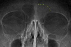
The examination is carried out mainly in direct or lateral projections, which allows assessing the volume and depth of the sinus, as well as identifying the presence of a pathological process, pathological substances in it. It is imperative to make sure that the sinus is not inflamed and that there is no purulent or other exudate in it. This is due to the fact that the frontal sinus is connected to the brain through the eye socket, accordingly, if there is an infection, it can quickly spread to the brain, causing various infectious diseases, including meningitis.
Hypoplasia of the left frontal sinus
This term means that the left frontal sinus is underdeveloped. At the same time, the right one is fully developed. Usually, the sinus begins to develop, then for some reason it slows down or completely stops developing. Often, this pathology does not manifest itself in any way, is completely asymptomatic, and does not cause any discomfort to the patient. It can be diagnosed during an examination. It is quite easily detected by percussion and correct palpation, causing painful sensations.
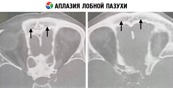
Aplasia of the left frontal sinus
Often, aplasia is a hereditary pathology and means a complete absence of the frontal sinuses, their underdevelopment. The pathology is formed when the process of normal formation of various cranial sections is disrupted. First of all, the facial surface of the brain is incorrectly formed.
It is often manifested by a slight depression or sinking of the frontal lobe of the head. At the same time, there is a complete or partial narrowing of other paranasal sinuses and the nasal canal. There is excess pressure on the facial or nasal wall, slight asymmetry. In the area of the canine fossa, a slight depression may be observed. It ends with a complete fusion of the nasal and facial walls.
Aplasia of the right frontal sinus
Unilateral pathologies develop quite often. In this case, facial asymmetry is well developed. The main symptom is also the insufficient development of the opposite sinus. When trying to puncture using a puncture, the needle immediately enters the soft tissues of the cheek. Most often found in men. Often causes sinusitis, affects the frequency of nasal pathologies. Pain is usually heard only during palpation or percussion.
Complications and consequences
The disease is completely asymptomatic in many people, and does not cause any consequences or complications. Usually, aplasia does not cause any inconvenience to a person. Whereas hypoplasia can lead to some complications. For example, underdeveloped sinuses can be complicated by sinusitis, otitis, and other inflammatory and exudative processes. The frontal sinus is connected to other paranasal sinuses, the nasopharynx, the ear, and the nasolacrimal canal by various channels. As a result, the existing infection can persist in these channels as a single system, transmitting the infectious and inflammatory process to any of the areas.
The danger is that the frontal sinus is connected to the brain via the bottom of the orbit. Accordingly, the inflammation can be transmitted to the brain. Also, if the bones are thin and porous, the infection can penetrate into the brain sections, causing inflammation of the meninges.
Externally, severe swelling and redness may appear, which are transmitted and spread to other sinuses and body parts. The danger is that the entire system may be affected. In this case, the infection can spread to the lungs, bronchi, trachea, causing corresponding inflammatory reactions. It can affect the eye, contributing to the development of the inflammatory process. Most often, conjunctivitis develops, vision is impaired, and lacrimation appears.
The danger lies in the accumulation of infection, which is accompanied by general weakness, high temperature, decreased attentiveness and performance. Pus, purulent-mucous exudate, which is capable of further spreading to neighboring areas, especially to the brain, can form, which can have extremely negative consequences.
The presence of pus in the sinuses is also dangerous, since the channel that connects the nasopharynx to the sinuses is very thin and can easily become clogged with purulent masses. Also, with the presence of pus, the mucous membrane increases, which makes the channel even narrower. Thus, the removal of pus to the outside will be disrupted, and surgery may be required. It is important to carry it out in a timely manner to prevent pus from entering the meninges.
[ 8 ]
Diagnostics hypoplasia and aplasia of the frontal sinuses.
Diagnosing malformations of the paranasal sinuses is usually not difficult. The diagnosis can be made based on a survey and visual examination of the patient, since the clinical picture is quite pronounced and specific. A standard physical examination is carried out using clinical research methods. Percussion can reveal a characteristic sound that will indicate the development of hypoplasia or aplasia. Palpation can be used to feel the frontal sinus, determine its border and volume. Auscultation is rarely used, since in this case it is not very informative.
If there is insufficient information to confirm the diagnosis, special laboratory and instrumental studies may be prescribed. Differential diagnostics are performed if several diseases have a similar clinical picture and make it difficult to differentiate.
Tests
Standard tests are prescribed: blood and urine tests. They allow identifying such disorders in the body as inflammatory or infectious processes, allergic or parasitic reactions. Inflammation and infection will be indicated by an increase in ESR in the blood, a shift in the leukocyte formula to the left, the presence of a large number of neutrophils, leukocytes, lymphocytes. The presence of an allergy will be indicated by a high level of eosinophils, basophils, and an increase in histamine in the blood. With a parasitic infection, an increased level of eosinophils will also be observed.
A bacteriological examination may be required if there is inflammation and it is necessary to determine the pathogen and select the optimal dosage of the drug. If a viral infection is suspected, virological and bacteriological studies are carried out. If an allergic reaction is suspected, allergological tests and an analysis for immunoglobulin E are carried out, which is the main indicator of allergy in the body.
Instrumental diagnostics
To conduct the study, the radiography method is used, which allows viewing the main sinuses of the nose in various projections, including the frontal, to identify possible foci of infection, signs of inflammation, bone defects. It is possible to differentiate hypoplasia from complete aplasia, to determine on which side the pathology is located.
An equally informative method is microrhinoscopy, in which the nasal cavity is probed with rubber catheters or metal probes. The study makes it possible to assess the condition of various sinuses, nasal passages, and also to determine the degree of underdevelopment of the sinuses, or to diagnose their complete absence. It is performed under local anesthesia.
The most informative method is considered to be computed tomography, with the help of which it is possible to comprehensively assess the condition of the nasal cavity and paranasal sinuses, identify possible anomalies and congenital defects, assess the degree of pathology, consider the presence or absence of an inflammatory process, a source of infection. Various tumors can be detected at an early stage of their formation. Allows to assess not only the condition of the skeletal system, but also soft tissues.
If necessary, fibrorhinoscopy is performed, which, together with microrhinoscopy, makes it possible to assess the condition of the microstructures of the nose and identify abnormally altered areas.
Differential diagnosis
Another important stage of diagnostics is conducting a medical genetic consultation. It includes a thorough analysis of family and hereditary anamnesis, which allows establishing an accurate diagnosis and comprehensively studying the causes and pathogenesis of the disease. During the consultation, concomitant factors are established, internal and external teratogenic factors that may affect the fetus are examined.
It is important to differentiate hereditary and non-hereditary diseases, as well as to determine the type of inheritance in each family, based on clinical and genealogical research methods. The goal is to determine the probability of the appearance of a patient with a genetically determined pathology in the family. It is important to choose the optimal method of treatment and rehabilitation as quickly as possible.
Treatment hypoplasia and aplasia of the frontal sinuses.
Treatment is used if the pathology causes discomfort to the patient. In the absence of any complaints, treatment may not be carried out. In the presence of pain, discomfort, difficulty breathing, inflammation, a conservative method of therapy is used, mainly the drug route is chosen.
Various medications are used, in particular, vasoconstrictor drops, sprays, solutions for rinsing the nasopharynx, mouth. Antihistamines are used for allergies and swelling. Mucolytic agents are used to stimulate the outflow of sinus contents and restore mucociliary clearance. Antibiotics, antiviral drugs, antimycotics, and immunomodulators may be prescribed as indicated.
Vitamin therapy is often performed. Physiotherapy procedures are performed if necessary. Physiotherapy procedures are most often used after punctures, releasing the sinus from purulent contents, which helps prevent relapses. In this case, warming up and UHF therapy are usually required.
Therapy helps reduce atrophic processes in the mucous membrane and prevent the development of inflammation. In some cases, it is even possible to prevent pathological changes in bone tissue. Physiotherapy is not used in cases of severe allergic reactions, as it can only worsen the pathology by increasing swelling.
It is recommended to conduct complex therapy, which will also include folk remedies, homeopathic preparations, medicinal herbs. You can do steam inhalations at home using various herbal decoctions, essential oils. Inhalations are contraindicated in the presence of pus, as this can cause complications. Also, various warmings are carried out, compresses, rinsing, rinsing are done. Massage and manual therapy are excellent treatments.
Hormonal and other means are used to relieve swelling and inflammation. Adrenalization of the mucous membrane has proven itself well. For this, frequent and abundant lubrication or irrigation of the mucous membrane with preparations containing adrenaline is performed. Similar preparations can also be used for instillation into the nose. Such therapy helps to reduce the thickness and looseness of the mucous membrane, respectively, inflammation decreases and excessive mucus production ceases.
Surgical methods are rarely used, only when conservative therapy is ineffective. Trepanopuncture is performed, during which the frontal sinus is punctured to clear it of accumulated transudate or exudate.
Prevention
Prevention is based on early detection of various inflammatory processes and anomalies. It is important to conduct timely medical and genetic counseling in order to promptly identify possible anomalies and develop a plan for further rehabilitation and treatment.
It is also important to maintain nasal hygiene, maintain immunity at a high level, avoid colds and other diseases. When the nose is stuffy, you should not blow your nose too hard, since the resulting mucus from the nasopharynx can get into the frontal sinuses through the channels and cause inflammation or blockage.
Prevention also includes hardening, physical exercise, proper breathing, and relaxation practices.
[ 15 ]
Forecast
If you see a doctor in a timely manner and undergo the necessary treatment, the prognosis is quite favorable. It can be unfavorable if an infectious and inflammatory process develops and there is no treatment. The greatest danger is the penetration of infection and pus into the meninges. Hypoplasia and aplasia of the frontal sinuses can be detected during medical and genetic counseling when planning a pregnancy.