Medical expert of the article

New publications
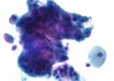
Highly differentiated adenocarcinoma
Last reviewed: 05.07.2025

All iLive content is medically reviewed or fact checked to ensure as much factual accuracy as possible.
We have strict sourcing guidelines and only link to reputable media sites, academic research institutions and, whenever possible, medically peer reviewed studies. Note that the numbers in parentheses ([1], [2], etc.) are clickable links to these studies.
If you feel that any of our content is inaccurate, out-of-date, or otherwise questionable, please select it and press Ctrl + Enter.
According to the degree of formation of a specialized phenotype during cell division during morphogenesis, such tumors are divided into several types, one of which is highly differentiated adenocarcinoma, marked by a high degree of differentiation and insignificant cell polymorphism.
That is, in this case the cell structure changes insignificantly, only the growth of the cell nucleus size is observed. The area of localization of this pathology is quite extensive.
Adenocarcinoma is a cancerous neoplasm that forms from the epithelium of glandular tissues due to a failure of their reproductive program.
 [
[ Causes of well-differentiated adenocarcinoma
Scientists and doctors have been trying to find the causes of highly differentiated adenocarcinoma for a long time, and so far with little progress. Once this is achieved, we can safely say that a medicine capable of ridding a person of this problem will soon be found, but for now medicine has learned to diagnose the disease, assuming the causes of its occurrence.
- Genetic inheritance.
- Frequent neuroses and stressful conditions.
- Age. Older people are more prone to this.
- Lack of balanced and varied nutrition. Low content of plant products in food. Fats and carbohydrates in the form of flour and sweet dishes prevail in food. Cancer, in some cases, can be provoked by an unsuccessful diet.
- Diabetes mellitus.
- Professional activities involving work with hazardous substances.
- Medical preparations.
- Highly differentiated adenocarcinoma of the rectum may be triggered by anal sex.
- High degree of obesity.
- Human papillomavirus infection.
- The cause of uterine cancer is said to be a hormonal imbalance caused by an increased amount of estrogen (female sex hormone) in the blood of a representative of the fair sex.
- Various types of diseases of vital organs: ovaries, rectum and colon, prostate gland.
But this list cannot be called final. It is difficult to voice all the risk factors that can provoke cancerous tumors of one or another organ.
Symptoms of well-differentiated adenocarcinoma
It is necessary to clarify that initially malignant tumors of such differentiation do not manifest themselves in any way and the patient does not even suspect its existence for a while. Only with time do symptoms of highly differentiated adenocarcinoma begin to appear. In this case, the symptoms may vary slightly depending on the location of the tumor.
- The appearance of aching pain in the area of the neoplasm.
- Rapid weight loss, loss of appetite.
- In case of bowel cancer, the following are observed:
- Bloody, mucous or purulent discharge in the stool.
- Frequent alternation of diarrhea and constipation.
- Intestinal bloating.
- Symptoms of uterine damage may include:
- Unpleasant smell.
- Atypical vaginal discharge between periods.
- A nagging pain symptom observed in the lower abdomen.
- Heavy discharge during menstruation.
- Pain caused by sexual intercourse.
Well differentiated adenocarcinoma of the colon
Colon cancer – this term often denotes several different forms of manifestation of a cancerous tumor, its histology and localization. This includes epithelial cancerous neoplasms of the rectum, colon, cecum and, actually, the colon.
Today, this pathology ranks second in the world, especially in highly developed industrial countries, in terms of incidence. In particular, the statistics provided by highly differentiated adenocarcinoma of the colon are quite deplorable: about 16 thousand patients diagnosed with colon cancer die annually in England and Wales alone. The United States voices an even more terrifying figure: new cases of the disease from 14 to 150 thousand people, while the annual mortality from this disease exceeds 50 thousand people.
The first symptoms that should alert a person and prompt him to seek advice from a doctor should be atypical discharge observed together with feces - these are streaks of blood (or even bleeding), mucous or purulent discharge.
By the color of the blood, a specialist can quite accurately guess even the localization of cancer: scarlet blood is typical of neoplasms of the rectum and anal canal, while dark blood is more typical for left-sided colon cancer. Most often, blood, mucus and feces are mixed, stating the greater reliability of the sign. Hidden bleeding is typical for right-sided colon lesions. Its manifestation may be weakness, pale skin and obvious signs of anemia.
Most often, problems with defecation are characteristic of severe late forms of the disease and are more common in malignant tumors of the rectum and left part of the colon. There are cases when colon cancer immediately manifests itself as acute intestinal obstruction. This situation requires immediate surgical solution.
Well-differentiated adenocarcinoma of the cecum
This is one of the most common cancer pathologies of the intestine. The maximum number of cases occurs in patients aged 50 to 60 years, but young people are not immune from it either.
The development of cancerous neoplasms can be triggered by:
- Villous or adenomatous polyps.
- Proctosigmoiditis is an inflammatory process occurring in the lower part of the sigmoid (colon) and rectum.
- Chronic proctitis.
It is practically impossible to foresee or predict this pathology. The main task of doctors and the patient himself is not to miss the symptoms and take adequate measures in time.
Well-differentiated adenocarcinoma of the sigmoid colon
Malignant neoplasms affecting the mucous membrane of the colon and rectum have become the scourge of modern society. In elderly people, this pathology ranks second in terms of disease intensity. The sigmoid colon is one of the sections of the intestine.
This is the degeneration of mucosal cells into cancerous formations, the localization of which is "chosen" to be the sigmoid colon. The pathology does not reveal any symptoms in the early stages of development, it can only be diagnosed through regular screening. In most cases, elderly people over 50 are at risk.
Well differentiated adenocarcinoma of the rectum
The "share" of malignant epithelial tumors of the rectum accounts for about 4 - 6% of cases of this "plague of the 20th century". The peak number of diseases is noted in highly developed countries such as the USA, Canada, Western European countries, Russia. A significantly smaller percentage is noted in African and Asian countries.
As a rule, highly differentiated adenocarcinoma of the rectum begins to manifest itself with the following symptoms:
- The patient does not lose the desire to go to the toilet; he almost always feels false urges to empty his bowels.
- Weakness is observed.
- The feeling of hunger comes less often.
- Decreased ability to work.
- There is a significant loss of body weight.
- Earthy complexion.
- Marked anemia.
- There is bloating and rumbling in the abdomen.
- Increased peristalsis.
- Constipation.
- As the tumor grows, anal bleeding appears, which intensifies over time, blood clots periodically come out, but there is no diffuse bleeding.
- At a later stage of the disease, hepatomegaly (pathological enlargement of the liver) and ascites (accumulation of free fluid in the peritoneal cavity (peritoneal dropsy)) are observed.
Symptoms of malignant neoplasms are largely determined by the size of the tumor, the level of invasion, and the location. As it grows, the symptoms become more pronounced and varied.
Doctors distinguish three stages of highly differentiated rectal adenocarcinoma:
- Stage I: neoplasm up to 2 cm in size, localized in the mucous and submucous layer of the rectum. No metastasis is observed.
- Stage II: the neoplasm is up to 5 cm in size, covers less than half of the intestinal lumen, localization – does not spread to nearby tissues.
- stage IIa – without metastasis.
- stage IIb – regional metastasis is observed.
- Stage III: the size of the cancerous area is over 5 cm, the area of overlap of the lumen of the rectum is more than 50%, deeper metastasis growth is observed.
Well differentiated gastric adenocarcinoma
Malignant neoplasm of the glandular epithelium of the stomach, that is, the development of oncology in the glandular layer of the stomach, is one of the most common oncological diseases in the world today. Stomach cancer ranks fourth among other types of cancer. A malignant tumor of the stomach can develop in any of its sections, but most often it is found in the antral and pyloric sections, that is, "at the exit" of the stomach.
The impetus for the progression of such a disease as highly differentiated gastric adenocarcinoma can be the Helicobacter pylori virus, chronic gastric ulcers, subtotal gastrectomy, atrophic gastritis and multiple other diseases of the gastrointestinal tract.
In this pathology, a mutation of the genetic apparatus of the affected cell is observed. It is quite difficult to diagnose this disease because for the time being, the aberrant cell is practically no different from the normal one. If an oncologist has diagnosed stomach cancer, then in 90% of cases this is already a severe stage of the disease, when it is quite difficult to help the patient. The probability of a fatal outcome is very high.
In addition to the above, the risk of developing highly differentiated gastric adenocarcinoma increases if the patient’s medical history includes:
- Adenomatous polyps.
- Problems with the integrity of the gastric mucosa epithelium.
- Menetrier's disease.
- With improper nutrition: abuse of smoked, oversalted foods, canned foods, products with preservatives, modified foods.
- Genetic inheritance.
- Overweight.
- Living or working in an area of high radiation.
In addition to the “traditional symptoms,” a malignant neoplasm in the stomach provokes:
- Changes in taste preferences.
- A feeling of heaviness in the stomach after eating.
- Non-infectious jaundice.
- Change in bowel movements.
- There is a loss of body weight, while the abdomen increases in volume.
- The appearance of pain and discomfort in the stomach area.
Chronic pancreatitis, smoking can provoke highly differentiated adenocarcinoma of the pancreas.
The malignant neoplasm of the stomach itself has many varieties, depending on the form of the tumor itself, the way it develops. An important histological characteristic of the tumor is the level of cell differentiation. If we are talking about highly differentiated adenocarcinoma, then the pathological cells do not differ much from the cells of the tissue that formed the tumor. Such a neoplasm develops relatively non-aggressively and has the most favorable prognosis against the background of tumors with a lower level of cell differentiation. However, other characteristics of highly differentiated gastric adenocarcinoma are also important for adequate treatment.
One of the most widespread and used classifications in the world is the Bormann classification, which identifies four main types of malignant neoplasms of the glandular epithelium of the stomach:
- Polypoid
This type of cancer has fairly clear boundaries, no ulcers. It is quite rare - about 6% of cases of malignant neoplasm of the stomach.
- Non-infiltrative (saucer-shaped)
This type of cancer resembles an ulcer in appearance and has more extended borders. If it occurs, a thorough histological examination is required to clarify the diagnosis.
- Infiltrative
This type of cancer tends to grow into deeper layers of the stomach walls, has no clear boundaries, and also resembles a stomach ulcer. This type of cancer tends to actively metastasize.
- Diffuse infiltrative (solid)
In this case, cancer grows into the deep layers of the stomach, the motor activity of the stomach is significantly reduced. If the cancer has developed extensively, then the stomach itself practically loses its functionality and narrows significantly. Ulcers, erosions and hemorrhages can be observed at the site of the lesion. In this type of cancer, oncological and infectious processes are often associated.
Among the listed types of cancer, the last two have the most unfavorable prognosis. They affect the stomach to the greatest extent and are quite difficult to diagnose in the early stages. Also, the last two types of malignant neoplasm of the glandular epithelium of the stomach have a higher tendency to metastasize, which significantly complicates the patient's treatment and worsens the prognosis.
Well differentiated adenocarcinoma of the prostate gland
Prostate cancer is a disease that mainly affects elderly men and is characterized by a mutation of the glandular epithelium cells of the alveolar-tubular structures. The predominant localization of the pathology is the peripheral region of the prostate gland. One of the modifications of such malignant neoplasms is highly differentiated adenocarcinoma of the prostate gland.
Malignant neoplasm of the glandular epithelium of the prostate gland is a malignant tumor formed from the glandular tissues of the prostate gland.
Today, prostate adenocarcinoma ranks first among malignant tumors in men. Highly differentiated adenocarcinoma is the least aggressive, but nevertheless, the danger of this disease is very high.
Most often, this disease occurs in older men, but over the years, younger men are increasingly faced with this problem. And on average, prostate cancer reduces the life expectancy of patients by 10 years.
The symptoms of this lesion, as with other types of cancer, begin to appear only in the later stages of the disease, when the obstruction begins to affect the ureters. Therefore, at earlier stages, this pathology can only be diagnosed during a doctor's examination. The diagnosis can be assumed by conducting a digital rectal examination by a proctologist. Then the PSA value is monitored and a biopsy is performed.
The reasons for this failure in the body are called:
- Age of the man.
- Equilibrium balance of nutrients.
- XMRV virus.
- Poisoning of a man's body with cadmium, or prolonged exposure to this substance.
Main symptoms:
- Manifestation of painful sensations in the hip joints. There is a feeling that the spine and ribs hurt.
- The feeling of weakness and apathy increases.
- An increase in the frequency and duration of urination is recorded, and the procedure becomes painful.
- Urinary incontinence may occur.
All these symptoms are also inherent to prostate adenoma, which confuses an inexperienced doctor in making the correct diagnosis. If adequate treatment is carried out when the pathology has not yet had time to grow, the prognosis for patients diagnosed with prostate cancer is favorable in most cases.
Highly differentiated prostate adenocarcinoma is a malignant neoplasm that shortens the life of the stronger sex by at least 5-10 years. Difficulty with diagnosis at early stages significantly increases mortality, second only to lung cancer.
The danger of the disease is also that, like many other oncological processes, it does not have clearly defined symptoms. As this disease develops, symptoms such as frequent urge to urinate may be observed. There is a feeling of incomplete emptying of the bladder, the stream is intermittent, and difficulties and painful sensations during urination are possible.
Similar symptoms may appear in a number of diseases of the prostate gland and urinary organs, so if they appear, you should in any case consult a doctor to rule out prostate cancer.
Highly differentiated adenocarcinoma practically does not metastasize. But this process in the case of prostate adenocarcinoma has its own characteristics. The prostate gland itself has a capsule. When the tumor grows into neighboring tissues, the capsule limits growth. Thus, metastases most often penetrate the bottom of the bladder and seminal vesicles.
In addition, the tumor can spread through the lymphatic and blood channels. But in the case of highly differentiated adenocarcinoma, this probability is very small and amounts to about 10%.
Well differentiated adenocarcinoma of the lung
Highly differentiated adenocarcinoma is a type of cancer that can develop from glandular tissue in any organ where it is present, in this case, lung tissue. Its cells are structurally similar to the cells of the organ in which it formed.
Quite often, highly differentiated lung adenocarcinoma manifests itself by the production of mucous secretions. In this case, the structure of the mucus is represented by large cells with a large nucleus located in the basal region. In the tissue lumens, cancer cells and mucous masses are observed together (there are tumors in which mucous formations are absent).
Risk factors include:
- Long-term smoking.
- Passive smoking. A person does not smoke himself, but is in close contact with smokers for a long time. In this case, the risk of cancerous tumors in a non-smoker increases by 30%.
- Professional activity, the production costs of which include the inhalation of carcinogens.
- Lack of fruits and vegetables in the diet.
- Living or working in an area with high radiation.
- Chronic lung diseases:
- Tuberculosis.
- Bronchitis.
- Pneumonia.
- Pneumonia.
Highly differentiated lung adenocarcinoma progresses slowly, but already in the early stages of the disease it is intensively spread by blood vessels, late metastasis is observed. In the natural course of the disease, without undergoing a course of treatment, the outcome is one - death.
Lung cancer has a number of distinctive features. In particular, this type of cancer occurs in men more often than in women, can actively metastasize, and is characterized by active mucus secretion. Metastases are especially dangerous. In this case, they can spread not only to neighboring organs, but also to the brain, liver, bones, and adrenal glands. Malignant neoplasms of the glandular epithelium of the lung also grow quite quickly (the tumor size can double in six months). All possible lung cancers are conventionally divided into small cell and non-small cell. Adenocarcinoma is the most common type of cancer among non-small cell lung cancers.
Highly differentiated adenocarcinomas are divided into acinar and papillary forms. In the former, glandular structures with large cells predominate, in the latter, papillary structures. Both varieties tend to form mucus, and the tumor cells themselves contain large vacuoles with mucus. Cancer most often develops in the peripheral parts of the lung, and it is quite rare to find tumors of this kind on large bronchi.
Also, highly differentiated adenocarcinomas include bronchioalveolar cancer, which is dangerous because it develops asymptomatically and is most often discovered by accident.
Otherwise, the main symptom is abundant sputum. The tumor is detected by microscopic examination of mucus, as well as by X-ray examination.
Well differentiated adenocarcinoma of the mammary gland
The topic of breast cancer is on everyone's lips today. The relevance of this problem worldwide is beyond doubt. Today, every thirteenth woman over the age of 20 faces this problem.
One of the types of breast cancer is highly differentiated adenocarcinoma. This is the development of a tumor from the glandular part of the cells of the mammary gland. Such a tumor does not differ significantly in structure and cell functions from the tissue that formed it, and is even capable of maintaining producing functions.
The pathology under consideration is a cancerous neoplasm consisting of mutated cells of glandular epithelium, having a corresponding localization. If the cellular structure does not differ much from the norm, the structure of the neoplasm visually resembles the natural outline of the gland and does not manifest itself pathologically until it transitions to later, advanced forms, highly differentiated adenocarcinoma of the mammary gland is stated. Such pathology almost completely supports the functioning of the replaced glands.
In addition to genetic predisposition, hormonal imbalance and a burdened heredity, the risk of developing highly differentiated adenocarcinoma can be pushed by:
- Frequent chest injuries.
- Mastopathy of fibrous or cystic nature.
- Women who gave birth for the first time after the age of 30.
- Puberty in girls also began much earlier than normal.
- Infertility.
- Menopause period.
- A benign tumor can degenerate into a cancerous neoplasm.
- Significant doses of hormonal drugs were taken for the treatment of other diseases.
- Congenital anomalies in the structure of a woman's breast.
- Smoking and alcoholism.
- Improper nutrition.
Symptoms of well-differentiated adenocarcinoma of the mammary gland:
- When palpated, elastic seals of the spherical outline are determined.
- Inverted nipple.
- The shape of the mammary gland has undergone changes.
- Increase in the size of the axillary, subclavian and supraclavicular lymph nodes.
- There is discharge from the nipple.
- Change in skin color in the chest area.
- The mammary glands of the right and left breasts are located at different levels.
- Edema appears.
- In the later stages, painful symptoms appear.
Highly differentiated adenocarcinoma itself can differ in a number of features. Depending on the location of the tumor, ductal and lobular cancer are distinguished. To choose the treatment tactics, it is very important to correctly determine the form of cancer. Breast cancer can be papillary (the rarest and most dangerous form of the disease), inflammatory (in its manifestations resembles mastitis), medullary (the tumor is large, but does not grow into neighboring tissues), Paget's cancer (a disorder caused by a tumor of the areola and nipple) and ductal infiltrative (the most common form of the disease) are also distinguished.
In addition, there are several stages of disease development - from zero to fourth. Stage 0 describes a tumor that does not go beyond the boundaries of its origin, at stage 1 the tumor is small in size, but invasive and affects neighboring tissues, at stage 2 the axillary lymph nodes near the tumor are affected, stage 3 is divided into two subgroups, in case of 3A the tumor is more than two centimeters, while the lymph nodes are fused, at stage 3B the tumor already grows into neighboring tissues and the skin of the chest, at stage 4 the tumor grows beyond the chest and can affect other organs, such as the liver, bones, lungs and brain.
Early diagnosis and adequate treatment can significantly improve a woman’s quality of life and prolong her life.
Diagnosis of well-differentiated adenocarcinoma
Any cancer diagnosis is a series of standard methods. Naturally, some differences still exist.
Diagnosis of well-differentiated adenocarcinoma includes:
- Analysis of patient complaints.
- Studying his medical history.
- Examination by a specialist.
- Clinical studies: a complete blood count, urine and stool analysis for occult blood, and other studies necessary to reconstruct a complete clinical picture.
- Hysteroscopy with biopsy. Conducting a histological examination of scraping materials (cytological smear) (in case of uterine cancer) or tissues of the diseased organ.
- Ultrasound examination of the "questionable" area.
- Digital rectal examination (if there is a suspicion of cancer in this area).
- Colonoscopy. An endoscopist has the ability to examine the condition of the mucous membrane of the inner layer of the colon. A special probe helps him make an assessment.
- Irrigoscopy (if colonoscopy did not provide a complete answer to all questions) is an X-ray examination of the colon with retrograde administration of a radiopaque agent into it.
- Endorectal ultrasound examination.
- If necessary, an X-ray is prescribed.
Who to contact?
Treatment of well-differentiated adenocarcinoma
Cancerous tumors of various localizations provide their own features of stopping the process. But in any case, complex treatment of highly differentiated adenocarcinoma is carried out. The use of several methods at once is practiced. The intensity of treatment is adjusted depending on the location of the lesion, the stage of tumor development and the presence or absence of metastases.
It is necessary to establish the pathology and carry out the necessary treatment in a short time, since in the case of a cancerous tumor, even a slight delay can cost the patient his life.
It is almost impossible to avoid surgical intervention, but modern methods allow, for example, in the case of highly differentiated rectal adenocarcinoma, to perform surgical treatment without opening the patient. But the result of therapy will be favorable only if healthy tissues located close to the pathology are protected from damage. To solve this problem, along with surgical intervention, radiation therapy is used. Radioactive cesium is also used. Its effect makes it possible to reduce the volume of the neoplasm.
To "destroy" mutated cells, oncologists actively prescribe chemotherapy. When conducting it, such drugs as Cisplatin (Platinol), Carboplatin (Paraplatin), Docetaxel (intensively prescribed in case of lung tumor diagnosis), Adriamycin, Bleomycin, Vinblastine, Fluorocyl and Epirubicin (in case of malignant pathology of the stomach and intestines) are often used.
Cisplatin (Platinol). The drug is used in the form of droppers or injections, into a vein. The dosage is set individually at the rate of 30 mg per m2 ( patient body surface). The drug is administered once every seven days:
- For a single application, every three to five weeks, the amount is calculated as 60 to 150 mg per m2.
- for daily use, a dosage of 20 mg/ m2 is used. The introduction is carried out over five days. Repeat the course after four weeks;
- The calculated amount of 50 mg per m2 of the patient's body area is administered every first and eighth day of a four-week block.
In combination with radiation exposure, the drug is administered intravenously daily, at a dose of up to 100 mg.
Depending on the tumor location, the oncologist may prescribe the drug intraperitoneally and intrapleurally. The amount of the drug delivered is determined by the doctor individually within 40 - 100 mg. If the drug is delivered directly to the tumor, Cisplatin is not diluted strongly.
The most common side effects are:
- Weakening of hair follicles and hair loss.
- Peripheral nerve neuropathy.
- Formation of ulcers in the oral cavity.
- Malfunction of the digestive organs.
- Nausea leading to vomiting.
- Depressive state.
- Apathy.
- Loss of appetite.
- Decreased vitality.
- Defect of taste.
- Anemia.
- Decreased number of platelets in the blood.
- Blocking immunity.
- There is a deviation from the natural color, structure of the skin and nails.
Docetaxel. The drug is prescribed intravenously. It is administered slowly over the course of an hour. A single dose is 75–100 mg/m2. The drip is administered once every three weeks.
All drugs used in chemotherapy are quite aggressive and their use is not without consequences for the body, which, in response to aggression, manifests itself in side effects. In order to partially or completely remove them, the oncologist has to prescribe additional drugs to the patient, which are designed to reduce these consequences.
Fluorocil. The drug is often used in treatment schedules. It is administered intravenously by drip. An oncologist prescribes it when the leukocyte count is critical. Fluorocil is a supportive agent. The daily dose of the drug is 1 g per 1 m2 of body area. The duration of administration is from 100 to 120 hours.
There is another protocol for taking it: 600 mg/m2. The drip is administered every first and eighth day of the month. If the drug is taken together with calcium, the dosage is reduced to 500 mg per m2. The drug is administered daily for three to five days. Then a four-week break is taken.
The course of treatment and rehabilitation time often lasts for six months or even more.
More information of the treatment
Prevention of well-differentiated adenocarcinoma
There are no specific preventive measures that can guarantee protection for yourself and your loved ones from malignant neoplasms of various localizations.
Prevention of highly differentiated adenocarcinoma, recommended by oncologists, is, first of all, taking steps to reduce the risk of developing the disease.
- Control your weight. Excess weight and thinness increase the risk of pathology.
- Proper nutrition.
- Active lifestyle.
- Moderate physical activity.
- Scheduled examinations by specialists.
- Adequate treatment of chronic diseases.
- Eliminate smoking, drugs and alcohol from your life.
- Walking in the fresh air.
- Learn to avoid stressful situations.
- A harmonious combination of exercise and rest.
- Minimize contact with harmful substances.
Prognosis of well-differentiated adenocarcinoma
In medicine, there is a term - five-year survival. The probability that a patient will be able to cross this Rubicon is influenced by several factors: the size of the tumor, the depth of its penetration into the affected organ and the presence of metastases.
The larger the tumor size and the deeper it has penetrated into the body tissues, the less optimistic the prognosis for highly differentiated adenocarcinoma. The presence of metastasis does not inspire optimism either. But the typical affiliation of a cancerous neoplasm to highly differentiated adenocarcinoma inspires optimism, since it is amenable to more effective treatment (unlike moderate or poorly differentiated).
The prognosis of highly differentiated adenocarcinoma is especially favorable when the pathology is diagnosed at an early stage of the lesion. Therefore, timely diagnosis and mobile adequate treatment are important. For example, the "five-year survival rate" for uterine cancer gives the following percentages:
- course of treatment when diagnosed at stage I - 86–98%,
- treatment when stage II is established - 70–71%,
- “five-year survival rate” in case of diagnosis of stage III is 32.1%,
- at stage IV - 5.3%.
Prognosis of well-differentiated adenocarcinoma of the colon
Due to the fact that highly differentiated cancer is effectively treated, the prognosis is more favorable than in the case of moderate or poorly differentiated cancer. But the treatment outcome largely depends on the stage of the tumor process. If it was diagnosed at an early stage of development, the survival prognosis is 90%. But the more the process progresses, the more favorable the prognosis becomes.
If the lymphatic system is already involved in the process, the percentage drops to 50. A tumor located on the right side of the colon gives no more than 20% survival.
Statistics show that the average time for relapses to occur is between one and one and a half years.
Highly differentiated adenocarcinoma is an insidious and dangerous disease, and your life largely depends on how attentive you are to your body, how well you learn to "read" its signals for help. Therefore, at the slightest discomfort, you should consult a doctor. It is better to be on the safe side than to miss a disease.