Medical expert of the article

New publications
Hemianopsia: types, effective medications
Last reviewed: 12.07.2025

All iLive content is medically reviewed or fact checked to ensure as much factual accuracy as possible.
We have strict sourcing guidelines and only link to reputable media sites, academic research institutions and, whenever possible, medically peer reviewed studies. Note that the numbers in parentheses ([1], [2], etc.) are clickable links to these studies.
If you feel that any of our content is inaccurate, out-of-date, or otherwise questionable, please select it and press Ctrl + Enter.
Epidemiology
Hemianopsia can be congenital or acquired. The acquired form is the result of damage to various brain structures. The congenital form often develops in the presence of other CNS pathologies (hemianopsia with an isolated course is an extremely rare disorder).
In the overwhelming majority of cases of hemianopsia, the disorder is found in the occipital lobe of the brain, a little less often in the parietal and temporal lobe, and very rarely in the visual apparatus and lateral geniculate body.
The acquired form is more often diagnosed in female patients.
Hemianopsia can occur in people of any age, but is most often diagnosed in people aged 30–50 years.
Causes hemianopsias
The causes of hemianopsia development can be congenital or acquired.
Congenital hemianopsia develops against the background of various failures in the intrauterine development of the fetus, or as a result of disturbances in the course of pregnancy - for example, with severe oxygen deficiency, with damage by viruses or bacteria, with organic pathology of the central nervous system, etc.
The acquired form of the disease can develop as a result of the following painful processes:
- basal form of meningitis;
- infectious inflammations in the form of meningoencephalitis, encephalitis;
- purulent inflammation, brain abscess;
- arterial aneurysms;
- disorder of blood flow in the brain after mechanical damage (trauma, wounds, concussion);
- oncology affecting the pituitary gland, posterior, frontal and temporal lobes of the brain;
- hypertension;
- intracranial hypertension;
- increased thrombus formation in the retinal vessels;
- damage to the myelin sheath of nerves;
- trophic disorder, extreme exhaustion of the body.
Hemianopsia does not rule out vascular causes, rather the opposite: the condition of the brain vessels directly affects the development of visual pathology.
For example, hemianopsia often occurs during a stroke; if treatment is started in a timely manner, this disorder is transient in the post-stroke period.
Risk factors
The main risk factors for the development of hemianopsia are considered to be conditions that lead to impaired blood flow in the tissues of the brain, as well as intracranial tumor processes, injuries and wounds to the skull, purulent inflammatory processes, and aneurysms in the area of the base of the brain.
Pathogenesis
It is believed that the most common factor in the development of hemianopsia are neoplasms that arise in the sella turcica area, as well as vascular aneurysms of the circle of Willis, TBI, and optochiasmal inflammation of the arachnoid membrane.
The visual system is damaged by craniopharyngiomas, tumors in the pituitary gland, tumor processes in the temporal lobe, aneurysms. The central neuron of the visual pathway and cortical visual centers are affected by tumors in the temporal or occipital lobes of the brain, TBI, aneurysms.
Hemianopsia develops as a result of organic disorders of the brain structures responsible for vision. It is considered the main topographic and diagnostic manifestation of damage to the visual pathway at different points, and primarily in the brain.
Symptoms hemianopsias
Hemianopsia manifests itself as a visual function disorder, which is accompanied by:
- headaches;
- semi-fainting and fainting states;
- increased sensitivity to light stimuli;
- paresthesia of the extremities.
The first signs depend on the cause of hemianopsia. For example, the "loss" of individual areas of the visual field can manifest itself in different ways. Thus, the outer halves of the visible image may disappear, or the inner ones, etc. Such features of symptoms are reflected in the classification of hemianopsia.
Scotomas and hemianopsias
When speaking about scotoma, we mean a dark zone in the visual field. This zone can be different – in the form of a circle, oval, triangle. It is formed in any part of the visual field and is not connected with its outlines.
Scotoma does not always manifest itself in the same way, so the following types are distinguished:
- positive appearance - explained by patients as a dark spot that occurs when there is damage to the vitreous body;
- negative type - appears as a result of damage to the path of vision, practically invisible to the patient;
- flickering appearance – explained by patients as a contour flickering that occurs against the background of migraine.
Scotomas occur on one side or both sides. Their appearance is mainly associated with the following reasons:
- pituitary neoplasms;
- damage to the myelin sheath of neurons;
- persistent high blood pressure;
- preeclampsia during pregnancy ;
- prolonged intoxication;
- severe exhaustion of the body;
- severe and frequent migraines;
- increased intracranial pressure;
- thrombosis of retinal vessels.
Scotomas are most often noticed by the patient himself, so the disease is detected at the very beginning of its development. The flickering type of pathology in the elderly may indicate the likelihood of an imminent stroke.
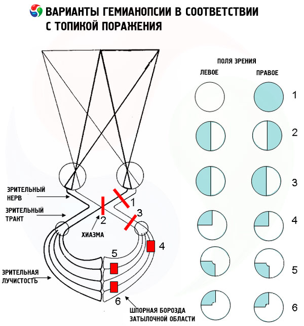
Forms
A pathology such as hemianopsia can proceed in different ways. For this reason, several types of this disease are distinguished.
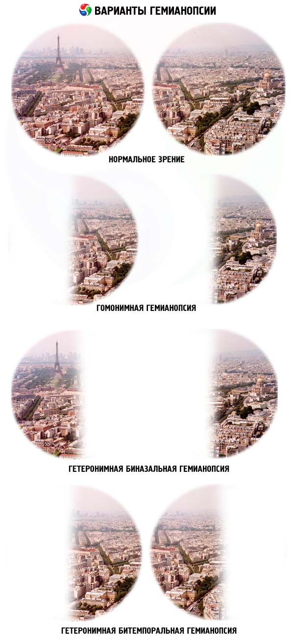
- Homonymous hemianopsia is a symmetrical loss of a pair of left or a pair of right halves of the visual field. For example, the patient is able to see only with the inner half of the left eye and the outer half of the right eye, or vice versa. By half we mean part of the visual picture.
- Heteronymous hemianopsia is the loss of a pair of external halves or a pair of internal halves of the visual field. Most often, the causes of this phenomenon are pathological changes in the cerebral cortex of the occipital lobe.
- Bitemporal hemianopsia is a heteronymic type of the disease with the loss of a pair of outer halves of the visual field.
- Right-sided hemianopsia is a type of homonymous type, when the patient perceives only the right half of the visual field. In this case, the boundary line dividing the perceived and lost halves coincides with the central vertical meridian.
- Left-sided hemianopsia is a homonymous disorder, opposite to right-sided hemianopsia. In this case, the patient perceives only the left half of the visual field.
- Binasal hemianopsia is a heteronymic type of the disease in which a pair of inner (nasal) halves of the visual field are lost.
- Contralateral hemianopsia is a homonymous type and is diagnosed when the occipital cortex is affected. This is often a consequence of a stroke. In some cases, this disorder is transient.
- The visual field is conventionally divided into quadrants for the convenience of diagnostic description of the disease. The term "quadrant hemianopsia" allows to accurately describe the localization of the dark spot that interferes with visual perception. Depending on the sector (quadrant) in which the spot is located, a distinction is made between lower quadrant and lower quadrant hemianopsia.
- Lower quadrant hemianopsia is characterized by damage to the area of the cerebral cortex, with superior localization relative to the calcarine groove.
- Upper quadrant hemianopsia develops when there is damage to the area of the cerebral cortex, with inferior localization relative to the calcarine groove of the temporo-occipital region.
- Partial hemianopsia is an incomplete loss of the visual field, on which spots of various sizes appear. As a rule, partial hemianopsia is observed at the initial stage of the pathology development.
- Bilateral hemianopsia, which is otherwise called bilateral, is characterized by the localization of visual impairment in two halves of the visual field.
- Tractus hemianopsia is a homonymous type of disease in which the pathological focus during diagnosis interrupts the arc of the pupillary reflex. In this case, diagnosis means determining the pupils' response to a light stimulus.
Complications and consequences
If you do not treat hemianopsia, do not carry out general or symptomatic measures, or treat the disease incorrectly, the pathology will gradually progress. Vision will deteriorate, the visual image will decrease in size.
The most common results of untreated hemianopsia are atrophic processes in the optic nerve, an increase in the size of the “blind” spot, and even complete loss of visual function.
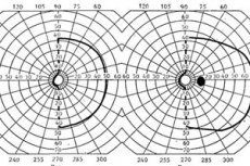
Diagnostics hemianopsias
Diagnostic measures for determining hemianopsia include procedures that evaluate the quality of visual function, the width of the visual field. If hemianopsia has just entered the first phase of development, then it is not always possible to detect it. The fact is that at the initial stage there are no pathological changes in the fundus, and the first signs appear about a year after the onset of the disease process.
Laboratory tests may be prescribed only to clarify the general state of health, to assess the functionality of the liver and kidneys, as well as the blood picture.
Instrumental diagnostics may include:
- computed tomography;
- radiography;
- carotid angiography;
- ultrasound;
- MRI of the brain.
If a tumor process is detected, the doctor may recommend taking a test to check the levels of certain hormones in the blood.
In addition, it is possible to conduct a special diagnostic test consisting of the following stages:
- The doctor and patient stand face to face, at a distance of about a meter.
- Both the doctor and the patient have one eye covered with a tight bandage.
- The patient directs his gaze towards the doctor's uncovered eye.
- The doctor moves the finger at an equal distance from the patient and from himself, starting from the periphery to the center. When the patient notices the finger in the visual field, he tells the doctor about it.
- Normally, the visual test results for the doctor and the patient should match. If the results differ, a visual impairment in the patient is suspected.
Differential diagnosis
Differential diagnostics of hemianopsia is carried out with such pathologies as ischemic neuropathy, glaucoma, retinal diseases. To clarify the diseases, the following studies are carried out:
- vasometry;
- ophthalmoscopy;
- tomographic studies, Dopplerography.
In some cases, a consultation with a neurologist or neurosurgeon may be necessary.
Who to contact?
Treatment hemianopsias
To eliminate hemianopsia and correct the visual field, it is necessary to completely eliminate the cause of the pathology. If this is not done, it will be impossible to cure hemianopsia, and further progression of the disease will lead to complete blindness. For example, in case of neurological pathologies, surgical treatment, chemotherapy may be prescribed - the choice of treatment depends on the type and severity of the problem.
Medicines for hemianopsia are practically not used, since there is no positive dynamics with conservative treatment. However, there are a number of drugs that are used to improve the patient's quality of life. Such drugs include, for example:
- Sumatriptan – used to relieve acute attacks of headaches and migraines, 1 tablet during an attack. No more than 2-3 tablets can be taken per day. Side effects – allergy, decreased blood pressure, chest and abdominal pain.
- Memoplant – used for vascular vision impairment, 1 tablet three times a day with food. Approximate duration of administration is 12 weeks. Side effects – nausea, allergy, stool instability.
- Cerebrolysin is prescribed for organic pathologies of the brain, post-stroke complications, and craniocerebral injuries. The drug is administered by injection: up to 5 ml as intramuscular injections, and up to 10-50 ml as intravenous injections. Side effects include: rare - tachycardia, pain at the injection site.
- Cerebroton – used for hemianopsia of vascular genesis, 1-2 tablets three times a day, regardless of food intake. Duration of administration – 1.5-2 months. Rare side effects – nausea, allergy.
Vitamins
With a varied and complete diet, there is no particular need to take additional vitamin preparations. In other cases, the doctor may prescribe special vitamin complexes as an addition to the main treatment of hemianopsia.
The following vitamin supplements are most often recommended for hemianopsia:
- Lutein complex – take one tablet 1-3 times a day.
- Optix is a vitamin-mineral complex preparation, taken one tablet daily for 3 months.
- Doppelherz Vitamins for eyes with lutein – allow to restore blood circulation and improve the quality of visual function. Taken daily for a long time.
- Focus forte – used daily for one and a half to two months.
Vitamins are used only in the context of the main therapy for hemianopsia, and they must be taken systematically over a long period of time.
Physiotherapy treatment
Physiotherapy and balneotherapy are primarily suitable for patients whose hemianopsia is a consequence of ischemic disturbance. In case of transient disturbances during periods of remission, general galvanization, galvanic collar, inductothermy, prolonged diathermy of the kidney and ankle zones, as well as UHF to the feet or solar plexus zone, and electrophoresis using the Vermel method are prescribed.
Patients with hemianopsia against the background of hypertension are given Ca-electrophoresis on the carotid sinus area, or a course of radon baths. The proposed procedures are best done every other day, and radon can be replaced with pine baths.
Patients whose hemianopsia is a consequence of dynamic circulatory disorders are allowed to use hydrogen sulfide baths simultaneously with oxygen therapy, as well as exercise therapy.
Contraindications to the appointment of physiotherapy for hemianopsia are diseases of the cardiovascular system (infarction, angina), circulatory disorders in the brain associated with an aneurysm or the third stage of hypertension.
Folk remedies
- In order to stabilize blood pressure and clear blood vessels in hemianopsia, use the following remedy. Wash and grind two oranges and two lemons, together with the peel, in a meat grinder. Add 2 tablespoons of honey to the resulting mass and keep the medicine for 24 hours at normal room temperature. Then pour the mass into a glass container and place in the refrigerator, taking 1 tablespoon three times a day. The duration of such treatment is not limited. You can wash down the remedy with warm unsweetened tea.
- Another popular remedy is used for hemianopsia. Grind five cloves of garlic, add the same amount of grated horseradish, pour the mass with dark sunflower oil. Place the mass in the refrigerator and take 1 teaspoon daily, along with 1 teaspoon of lemon juice, three times a day. The course of treatment can last from 4 to 12 weeks, then take a break for one month.
- A good effect in hemianopsia is achieved by treating it with golden mustache tincture. To prepare the tincture, cut off 35 “joints” of golden mustache, grind them, insist in medical alcohol for two weeks, filter. Use 1 tbsp. of tincture together with 1 tbsp. of dark sunflower oil. The medicine is taken 20 minutes before meals, three times a day. The course of treatment is 10 days, after which you need to take a break for 5 days. Then - another course of 10 days, but the next break should already be 10 days. So alternate five- and ten-day breaks until the medicine runs out.
 [ 36 ], [ 37 ], [ 38 ], [ 39 ]
Herbal treatment
- Hawthorn tincture is taken for a month, 25 drops per day. Then you should take a break for 2 weeks, and after that the course can be resumed.
- Prepare a decoction of 10 mulberry leaves and 500 ml of boiling water. Keep on low heat for 2 minutes, then leave to infuse for 20-30 minutes. Drink daily instead of tea. The course can last from 3 to 4 months. Mulberry helps stabilize blood pressure and normalizes blood vessels.
- Collect 12 medium cones in a pine forest, wash them, crush them and pour 500 ml of good vodka over them, leave for 2 weeks. Take 1 teaspoon with warm tea. The course of therapy lasts 7 days, after which a break is taken for one month.
- Prepare a mixture of 10 g of lemon balm, 10 g of speedwell, 30 g of strawberry leaves, 40 g of hawthorn flowers or berries. Take 1 tbsp of the resulting raw material and pour 300 ml of boiling water. Filter and drink instead of tea daily until you feel better. It is allowed to add honey to the warm drink.
Homeopathy
The decision to use homeopathic remedies for hemianopsia should be agreed upon with the doctor, after a thorough study of the clinical picture and conducting clarifying diagnostic studies. Depending on the cause, the following homeopathic remedies may be recommended:
- Aurum iodine, Barium carbonicum – improve blood circulation in the brain;
- Conium – normalizes vascular tone, especially relevant in the post-stroke period;
- Crategus, Arnica – have a positive effect on cerebral circulation;
- Ignatia amara - will help with hemianopsia, which occurs with headaches and high blood pressure;
- Cactus grandiflorus, Opium – stabilize blood pressure in hypertension;
- Staphysagria – improves vascular tone, normalizes vascular pressure.
A huge plus of homeopathy is the absence of side effects during treatment. However, specialists do not provide general recommendations on dosages: the dose is set individually, depending on the characteristics of the disease and the patient's constitution.
Surgical treatment
Surgical treatment for hemianopsia is indicated if it is caused by an oncological disease. Tumor removal, chemotherapy, and radiation therapy are performed.
Hemianopsia associated with traumatic brain injury may also require surgical intervention, which usually involves removing hematomas and suturing damaged tissue and vessels.
Prevention
Hemianopsia is an insidious disease. And, first of all, in the sense that at the initial stage of development it is almost impossible to detect hemianopsia. To protect yourself from troubles, it is advisable to have an annual examination by an ophthalmologist.
In addition, attention should be paid to a number of preventive measures:
- It is important to observe safety precautions in production, during physical activity, while driving a car, and in all other cases where there is an increased risk of head injury.
- If there are signs of a tumor process in the brain (seizures, vestibular disorders, strabismus), you must definitely consult a neurologist and go through all stages of diagnosis.
- In summer, it is necessary to take measures to prevent bites from encephalitis ticks.
- At any time of the year it is necessary to prevent the occurrence of infectious pathologies and strengthen the immune defense.
Forecast
Restoration of full visual image is possible, but the likelihood of such a positive outcome depends on many factors:
- from the complexity of the pathology that led to the development of hemianopsia;
- from the prescribed treatment;
- from the duration of visual impairment;
- from the stage of the pathological process;
- depending on the age and general health of the patient.
As a rule, recovery occurs within six months after the start of treatment, or does not occur at all. If the disease is neglected, or treatment was started late, then hemianopsia can end in partial or complete loss of vision.