Medical expert of the article
New publications
Hematocolpos
Last reviewed: 04.07.2025

All iLive content is medically reviewed or fact checked to ensure as much factual accuracy as possible.
We have strict sourcing guidelines and only link to reputable media sites, academic research institutions and, whenever possible, medically peer reviewed studies. Note that the numbers in parentheses ([1], [2], etc.) are clickable links to these studies.
If you feel that any of our content is inaccurate, out-of-date, or otherwise questionable, please select it and press Ctrl + Enter.

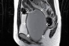
Gynecological problems include the accumulation of menstrual blood in the vagina – hematocolpos (from the Greek haima – blood, kolpos – vagina).
Epidemiology
There are no records of cases of accumulation of menstrual blood in the vagina, but cases of abnormalities of the genitourinary system in women make up just over 5% of the population.
Congenital defects in the form of atresia of the hymen are rare: one case per 2 thousand girls (according to other data, one case per 1000-10000 women), while this defect is the most common cause of vaginal obstruction of congenital origin.
The accuracy of statistics is questionable. Thus, according to some data, transvaginal (transverse vaginal) septum occurs in only one woman per 70 thousand; in other sources, the frequency of this anomaly is estimated at one case per 2-2.5 thousand women.
Causes hematocolpos
The main causes of hematocolpos are vaginal anomalies of a congenital nature: atresia of the hymen and transverse septum of the vagina – a connective tissue membrane. [ 1 ]
This condition can also be observed with a strong narrowing of the vaginal lumen (stricture) or its closure (atresia), which can be either congenital or acquired.
Acquired vaginal stricture or vaginal stenosis is associated with episiotomy (cutting the perineum and vaginal wall during childbirth), surgery for pelvic organ prolapse in women, and late effects of radiation therapy for uterine, cervical, vaginal, or colorectal cancer.
Risk factors
The risk of hematocolpos increases with developmental defects of the vagina and uterus, in particular, the above-mentioned congenital vaginal anomalies, which arise due to disturbances in the intrauterine development of the urogenital organs of the fetus. In the female fetus, they develop from mesodermal (primary) rudiments - the so-called Müllerian (paramesonephric) ducts. And due to their incomplete fusion, lack of fusion with the urogenital sinus, as well as incomplete involution of their remnants, organogenesis is disrupted.
The etiological factor of such disorders may be any teratogenic effect on the fetus in the first and early second trimester of pregnancy, as well as gestational diabetes.
In addition, vaginal anomalies can be part of genetically determined syndromes, such as Robinow syndrome (Robinow-Silverman-Smith syndrome), McKusick-Kaufman syndrome, and a rare congenital anomaly of the genitourinary system – Herlin-Werner-Wunderlich syndrome.
And congenital adrenal hyperplasia increases the risk of vaginal stenosis, accompanied by hematocolpos.
Pathogenesis
The pathogenesis is caused by the blockage of vaginal discharge (blood with a detached part of the mucous membrane of the uterus - the endometrium), which is removed from the uterine cavity with each menstruation.
Atresia of the hymen and hematocolpos are linked by a cause-and-effect relationship, since the continuous membrane, which has no natural perforation, that surrounds the opening of the vagina completely closes it and prevents the outflow of menstrual blood.
Symptoms hematocolpos
It should be borne in mind that the first signs of accumulation of menstrual blood in the vagina may appear only after menarche. That is, in the presence of congenital vaginal anomalies, hematocolpos appears in pubertal girls after the onset of menstruation.
In this case, the following symptoms are observed:
- cyclic pain with spasms in the suprapubic region;
- back pain (in the lumbar region) and intense pelvic pain with tenesmus (false urge to defecate);
- vomit;
- bloating, constipation or diarrhea;
- problems with urination (urinary retention).
Some women with vaginal stenosis associated with amenorrhea (absence of periods) may also have a painful mass in the abdominal area.
Hematocolpos and hematometra (hematometrocolpos) can be observed simultaneously – accumulation of menstrual blood in the uterine cavity: due to the same hymenal atresia or cervical canal stenosis. [ 2 ], [ 3 ]
Complications and consequences
The most likely complications and consequences of hematocolpos are:
- cryptomenorrhea (or retrograde menstruation with the absence of menstrual discharge from the vagina);
- accumulation of menstrual fluid in the fallopian tubes (hematosalpinx);
- endometriosis;
- recurrent urinary tract infection;
- hydronephrosis and obstructive acute renal failure (developing due to compression of the ureters);
- pelvic infections with abscess and peritonitis.
Diagnostics hematocolpos
For more details, see – Diagnosis of Vaginal and Uterine Malformations
Instrumental diagnostics are carried out using: transabdominal ultrasound of the pelvic organs and uterus; computed tomography or magnetic resonance imaging of the pelvic organs.
Differential diagnosis
Differential diagnosis includes dysmenorrhea of puberty, pelvic venous congestion syndrome with chronic pain, pyocolpos.
Treatment hematocolpos
Treatment of hematocolpos is surgical and, depending on the cause, may involve an incision in the hymen membrane (hymenotomy), complete hysterectomy, or removal of the vaginal septum (with access through the perineum).
More details in the publication – Treatment of malformations of the vagina and uterus.
Prevention
Measures to prevent congenital vaginal anomalies have not yet been developed.
Forecast
With intervention to eliminate the anatomical causes of hematocolpos and hematometra, the prognosis for the disease is favorable.

