Medical expert of the article

New publications
Holoprosencephaly in the fetus and newborn: causes and prognosis
Last reviewed: 04.07.2025

All iLive content is medically reviewed or fact checked to ensure as much factual accuracy as possible.
We have strict sourcing guidelines and only link to reputable media sites, academic research institutions and, whenever possible, medically peer reviewed studies. Note that the numbers in parentheses ([1], [2], etc.) are clickable links to these studies.
If you feel that any of our content is inaccurate, out-of-date, or otherwise questionable, please select it and press Ctrl + Enter.
Epidemiology
Holoprosencephaly is not considered a very common defect: according to statistics, the disease occurs in 0.06-0.2 newborns per 1 thousand. In case of spontaneous termination of pregnancy, up to 4 pathological cases per 1 thousand can be observed.
In the list of developmental defects of the central nervous system, holoprosencephaly is diagnosed in 2-3% of cases, as well as in approximately 5% of fetuses during autopsy (necropsy).
In more than half of patients, holoprosencephaly is isolated, and in about 22% of cases it occurs in combination with a large number of other developmental defects. In more than 17% of cases, a syndromic variant of the disease is diagnosed.
Holoprosencephaly is more often diagnosed in girls.
Causes holoprosencephaly
Holoprosencephaly is a congenital developmental disorder that occurs as a result of a failure in the mechanism of intrauterine growth of the fetus. The disorder is usually formed by autosomal recessive, autosomal dominant and X-linked inheritance.
One of the causes that is not related to chromosomal abnormalities is a genetic mutation. Currently, the following genes have been identified that are susceptible to mutation with subsequent development of holoprosencephaly:
- SIX3 2p21;
- TGIF 18p11.3;
- ZIC2 13q32;
- SHH 7q36.
Experts also draw attention to the negative impact of endocrine pathologies in the mother, smoking, drinking alcohol and taking medications from the salicylate series.
Risk factors
Factors in the development of holoprosencephaly are conventionally divided into hereditary and external.
A number of hereditary factors are chromosomal disorders that lead to changes in the karyotype:
- Orbeli syndrome;
- Ecardi syndrome;
- Meckel's syndrome;
- trisomy 13.
External factors of holoprosencephaly are:
- endocrine diseases in the expectant mother, insulin-dependent diabetes mellitus;
- low cholesterol;
- consumption of alcoholic beverages;
- taking certain medications (salicylates, methotrexate, retinoic acid, misoprostol, diphenylhydantoin).
Pathogenesis
Non-syndromic hereditary holoprosencephaly is transmitted most often by an autosomal dominant variant, but other inheritance routes are also possible. About 30-50% of pathologies have a clear connection with a chromosome disorder: a predetermined distribution of defects indicates the presence of at least twelve different loci.
The first mutated gene to be discovered is SHH, which is located at locus 7q36. Mutational changes in SHH cause approximately 35% of familial abnormalities with autosomal dominant holoprosencephaly.
The protein substance SHH is a signaling protein that is produced and is necessary for normal fetal development, both in animals and in arthropods.
Mutational changes in this protein substance lead to functional disorders – holoprosencephaly. Thus, certain cytogenetic defects that affect the process of transferring genetic information from DNA through RNA to proteins and polypeptides belong to translocations that extend to 15-256 kilobases at the 5'-end of the coded episode of the SHH gene. These types of chromosomal mutations are called positional, since they do not affect the change in the coding sequence, but disrupt control and change the structure of chromatin, which affects the process of transferring genetic information SHH.
Symptoms holoprosencephaly
Modern specialists distinguish several types of holoprosencephaly, differing from each other in symptoms, severity and frequency of occurrence. Geneticists still cannot pinpoint the exact reason why the disease manifests itself in one form or another. Presumably, the development of a certain type of holoprosencephaly is influenced by the location of the chromosome damage, the characteristics of the pregnancy, or some other factors.
Medicine knows the following types of holoprosencephaly:
- Alobar holoprosencephaly is the most complex type of this pathology, in which severe developmental defects are observed, affecting the brain, facial area, and other organs. Among all cases of the disease, this type occurs in 20% of cases.
- Semilobar holoprosencephaly is the most common type of the disorder, with complex but less defined developmental defects. Among all patients, the semilobar type is diagnosed in approximately 50% of cases.
- Lobar holoprosencephaly is the "mildest" type of the disease, which is amenable to relative surgical and drug correction. This type of pathology occurs in approximately 20% of patients with holoprosencephaly.
There is also a fourth type of holoprosencephaly, which began to be considered separately from other types only a little over twenty years ago. This is a rare type of mid-hemispheric fusion - it is characterized by blurred signs that differ from the classic course of the disease.
 [ 21 ], [ 22 ], [ 23 ], [ 24 ], [ 25 ], [ 26 ], [ 27 ]
First signs
Symptoms of holoprosencephaly can vary greatly, depending on the type of disease. Only a few early signs may be common:
- cleft palate and cleft lip;
- periodic attacks of convulsions;
- mental inadequacy;
- impaired reflexes;
- pathological changes in the cornea and retina.
- The alobar type of holoprosencephaly is characterized by cyclopism, underdevelopment of the nose, significant discrepancy in head size, and multiple defects in other organs. The development of the alobar type in the overwhelming majority of cases ends in spontaneous termination of pregnancy (or stillbirth): the few surviving babies die within the first six months of life.
- The semilobar type of holoprosencephaly also has characteristic symptoms: close placement of the eye sockets, a slight decrease in the head, and a violation of the development of the nasal passages. Infants with such defects die during the first 24 months of life.
- The lobar type is characterized by such signs as anomalies in the structure of the palate and upper lip. Children born with such pathology can live to an older age, provided that timely surgical intervention is performed.
- With average interhemispheric fusion, there are no defects in the child's face. However, mental retardation, seizures, and other neurological symptoms occur.
In most cases, fetal holoprosencephaly is detected during a woman's pregnancy or immediately after the baby is born. In addition to external manifestations, the patient is found to have endocrine dysfunction, renal dysplasia, damage to the respiratory system and other organs. Heart defects, autoimmune diseases, lack of reflexes, etc. are often diagnosed.
Complications and consequences
The consequences of holoprosencephaly generally depend on the degree of damage to the brain.
In alobar holoprosencephaly, a fatal outcome is most likely.
In other cases, children suffer not only from mental retardation, but also from seizures, problems with reflexes, and dysfunctional disorders of the brain stem.
According to statistics, more than 40% of children with holoprosencephaly do not survive beyond 5-6 months, about 80% die in the first year of life.
With the lobar type of the disease, a child can live for several years, but still dies from dysfunctional deficiencies.
Diagnostics holoprosencephaly
Any pediatrician or neonatologist can easily distinguish a healthy child from a baby with holoprosencephaly, since the manifestations of the disease are very specific.
Holoprosencephaly is often diagnosed in the first trimester of pregnancy based on the results of an ultrasound examination.
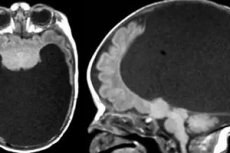
Holoprosencephaly on ultrasound is manifested by a sharply abnormal structure of the skull and brain of the unborn child:
- In the alobar type of holoprosencephaly, the brain has the appearance of a bubble with liquid contents, without the slightest signs of distinct hemispheres.
- In the semilobar type of the disease, ultrasound reveals a peculiar groove crossing the posterior part of the brain, which corresponds to a partial demarcation into hemispheres.
- In the lobar type of holoprosencephaly, diagnosis is somewhat difficult: symptoms of incomplete delineation of the brain are detected only in the depths of the organ, since the thalamus, corpus callosum and ventricles are predominantly affected.
Blood and urine tests are performed on women to detect diabetes in the expectant mother, which will allow us to suspect a developmental disorder of the fetus.
Instrumental diagnostics of holoprosencephaly in the prenatal period is the use of molecular genetic methods. Material for study in case of suspected congenital anomalies is taken in case of inadequate ultrasound examination indications, in case of previous cases of birth of children with various congenital pathologies, in case of certain signs of genetic problems in parents, in case of diabetes mellitus in the expectant mother. Amniocentesis or chorionic villus biopsy are used to collect material. Genetic tests often consist of direct separation of the SHH gene to determine disorders, or studying the karyotype of the future child to assess chromosomal inconsistencies. It is worth noting that in more than half of the cases the test does not detect a karyotype failure, which allows us to classify this method as uninformative procedures.
What do need to examine?
What tests are needed?
Differential diagnosis
Differential diagnostics of lobar holoprosencephaly is carried out with septo-optic dysplasia. Important distinguishing symptoms in this case are:
- hypoplastic changes in the frontal lobes;
- malformation of the temporal horns in the lateral ventricles;
- absence of frontal horns.
At the prenatal stage, holoprosencephaly should be differentiated from severe ventriculomegaly, encephalocele, cerebral cystic formations, and hydranencephaly.
Who to contact?
Treatment holoprosencephaly
Holoprosencephaly does not require any special treatment. If the doctor deems it appropriate, he or she may prescribe surgical treatment with correction of facial defects. Symptomatic treatment is also administered to alleviate the child's suffering.
Alobar and semilobar types of holoprosencephaly are not amenable to neurosurgical intervention, since the baby’s condition may not only not be corrected, but may also worsen.
The lobar type of the disease can be treated surgically: defects of the upper palate and lip are promptly eliminated, and adequate nasal passages are formed.
All forms of pathology require the use of anticonvulsant therapy, with possible individual correction of other disorders.
The doctor may prescribe the following medications for holoprosencephaly:
Dosage and method of administration |
Side effects |
Special instructions |
|
Suxilep |
Prescribed in individual dosages, approximately 5 mg per kg of weight. |
Dyskinesia, dizziness, weakness. |
The drug may have a myelotoxic effect. |
Sibazon |
Prescribed in individual dosages. |
Muscle weakness, hiccups, sleep disturbances. |
With prolonged use, drug dependence may develop. |
Mydocalm |
Prescribed at a rate of 5 mg per kg of weight, three times a day. |
Muscle weakness, low blood pressure. |
The drug is not prescribed to children under 3 years of age. |
Cerebrolysin |
Prescribed in individual dosages. |
Dyspepsia, tremor, feeling of heat, allergy. |
Injections of the drug are carried out slowly to avoid local side effects. |
Vitamins
Vitamins are not of decisive importance in alleviating the condition of a child with holoprosencephaly. However, in some cases, vitamin therapy in maintenance doses may be considered:
Type of vitamin |
Effect of vitamin |
Dosage |
Vitamin A |
Improves vision and the condition of mucous tissues. |
1250 IU |
Vitamin D |
Provides calcium and phosphorus exchange, improves bone tissue formation. |
300 IU |
Ascorbic acid |
Strengthens the immune system and blood vessels. |
30 mg |
Vitamin B 1 |
Improves the condition of the nervous system. |
0.3 mg |
Vitamin B 2 |
Ensures normal metabolic processes. |
0.4 mg |
Vitamin B 5 |
Responsible for hormonal balance and antibody production. |
2 mg |
Vitamin B 6 |
Responsible for the processes of hematopoiesis. |
0.5 mg |
Vitamin B 9 |
Responsible for the formation of new cellular structures. |
25 mcg |
Vitamin B 12 |
Improves the function of the nervous system. |
0.4 mcg |
Vitamin PP |
Responsible for digestion processes. |
5 mg |
Vitamin H |
Improves liver function. |
15 mcg |
Tocopherol |
Strengthens blood vessels. |
3 mg |
Vitamin K |
Normalizes blood clotting processes. |
10 mcg |
Physiotherapy treatment
Physiotherapy treatments generally do not provide significant results for any form of holoprosencephaly.
Folk remedies
It is difficult to talk about folk treatment for such a serious developmental abnormality of a child as holoprosencephaly. This disease is so serious that sick children in most cases do not live even six months - and only in rare cases can their existence be extended with the help of surgical treatment.
Herbal treatment in the form of individual folk recipes can only help symptomatically: reduce the severity of cramps, ease breathing and normalize the functioning of the baby's nervous system.
- Mix 1 part crushed wormwood seeds and 4 parts vegetable oil, leave overnight. Give the child 1-2 drops mixed with sugar.
- Brew 15 g of thyme in a glass of boiling water and offer the patient 1 tbsp. three times a day.
- The petals of the field poppy are dried, ground to a powder and boiled in milk. Honey is added and the patient is given a little during the day.
- Prepare a mixture of 1 teaspoon of anise seeds, 1 teaspoon of fennel seeds, 1 teaspoon of caraway seeds and 2 teaspoons of mint leaves. Brew one tablespoon of the mixture in 200 ml of boiling water, leave for 30 minutes, filter. Give the patient to drink in small quantities throughout the day.
- Brew 2 teaspoons of birch buds like tea. Drink 100 ml twice a day.
- Rub mustard oil into the patient's hands and feet.
- Add honey to food and drinks.
Plants such as lily of the valley, white mistletoe, valerian rhizome, walnut partitions, hawthorn and barberry fruits, hop cones, as well as oregano, thyme, heather, and sweet clover have a good anticonvulsant and tonic effect.
[ 39 ], [ 40 ], [ 41 ], [ 42 ], [ 43 ], [ 44 ]
Homeopathy
Homeopathic medicines for holoprosencephaly can only be prescribed by a doctor: if traditional medicines were prescribed at the same time, they cannot be cancelled.
As a rule, a 30-cent dilution is used: a grain is diluted in 100 ml of water, and the patient is given 1 teaspoon daily, half an hour before meals.
- Zincum metallicum - for childhood convulsions.
- Veratrum album - for stiff joints, muscle rigidity.
- Stramonium - for dementia and seizures.
- Stannum metallicum - for convulsions and spasms.
- Plumbum metallicum – for muscle spasms, neuroses.
- Moshus - for convulsions, loss of consciousness.
Psychological help for holoprosencephaly
Pregnancy is an important and responsible period in a woman's life. However, the diagnosis of "fetal holoprosencephaly", made almost at the very beginning of pregnancy, brings a lot of fears and worries to the expectant mother, and also puts her in front of a difficult choice: to decide on an abortion, or to give birth to a child (what if the diagnosis turns out to be wrong)? At such moments, a woman worries not only about the health of the baby developing in the womb, but also about whether the problem will recur in subsequent pregnancies, whether she will be able to have children at all, etc.
Sometimes the situation that has arisen frightens the pregnant woman so much that her relatives seriously begin to worry about her mental state. At such moments, it is better to consult a psychologist or psychotherapist. An experienced psychologist can provide support to a woman during such a difficult period, restore harmony and faith in the future. If necessary, psychological assistance can be provided to all family members.
Prevention
To prevent the development of holoprosencephaly in a baby, preventive measures are taken even during the period of pregnancy planning and conception. A woman who is preparing herself for the role of a mother should be very attentive to her health. Even before pregnancy, all existing diseases should be treated, a dentist, gynecologist, and geneticist should be consulted.
Both during the period of planning a baby and already during pregnancy, it is strictly forbidden to drink alcohol, smoke, take drugs, or take medications not prescribed by a doctor. The attending physician, in turn, must know that the woman is planning a pregnancy: based on this, he prescribes medications that will not affect the quality of conception and the formation of defects in the fetus.
A pregnant woman, starting from the first days of pregnancy, must protect her health, not allow the influence of stress and excessive loads. It is also important to register with a women's consultation clinic in a timely manner, take the necessary tests and listen to the recommendations of a medical specialist.
Forecast
Holoprosencephaly has an extremely unfavorable prognosis, the degree of which depends on the severity of the detected defects. The mortality rate depends on the type of disease. In a large number of women diagnosed with fetal holoprosencephaly, pregnancy ends in spontaneous termination - thus, the human body itself decides that the future baby does not have the ability to exist. Among babies born with holoprosencephaly, most die during the first year of life. With moderate developmental defects, there is a chance that the patient will survive the infancy stage and live to adolescence.