Medical expert of the article

New publications
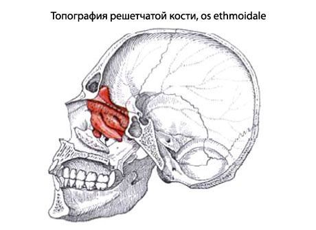
Lattice bone
Last reviewed: 06.07.2025

All iLive content is medically reviewed or fact checked to ensure as much factual accuracy as possible.
We have strict sourcing guidelines and only link to reputable media sites, academic research institutions and, whenever possible, medically peer reviewed studies. Note that the numbers in parentheses ([1], [2], etc.) are clickable links to these studies.
If you feel that any of our content is inaccurate, out-of-date, or otherwise questionable, please select it and press Ctrl + Enter.
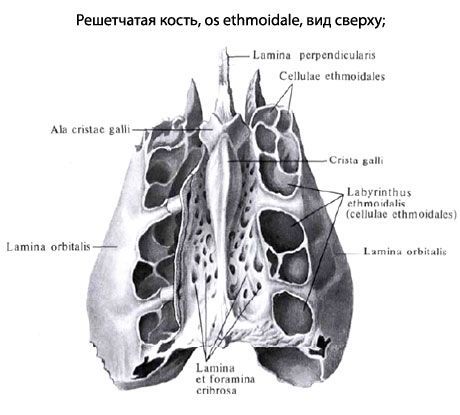
Ethmoid bone(os ethmoidalis) is part of the facial skull, forming, together with other bones, the walls of the nasal cavity and the orbit. The ethmoid bone has a horizontally located ethmoid plate, from which a perpendicular plate extends downwards into the nasal cavity. On the sides, to the right and left of the perpendicular plate, are the ethmoid labyrinths.
The cribriform plate (lamina cribrosa), which occupies the upper part of the bone of the same name, has numerous openings for the fibers of the olfactory nerve. Above the cribriform plate, the cock's comb (crista galli) projects upward along the midline. Anterior to the comb is the blind opening, in the formation of which the frontal bone participates.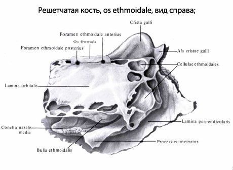
The perpendicular plate (lamina perpendicularis), located in the sagittal plane, participates in the formation of the upper part of the nasal septum.
The ethmoid labyrinth (labyrinthitis ethmoidalis) is attached to the perpendicular plate at the top on the right and left. The labyrinth is formed by air-filled bone ethmoid cells (cellulae ethmoidales). On the medial side of the ethmoid labyrinth there are curved bone plates - the superior and middle nasal conchae (conchae nasales superior et media). Due to these conchae, the surface of the mucous membrane covering them increases.
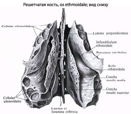
Between the superior and middle conchae is the narrow superior nasal meatus (meatus nasi superior), and under the middle nasal meatus is the middle nasal meatus (meatus nasi medius). From the middle nasal conchae extends downward and laterally the uncinate process (processus uncinatus). Behind this process the ethmoid vesicle (bulla ethmoidalis) protrudes from the wall of the middle nasal conchae into the middle nasal meatus.
Between the uncinate process in front and the ethmoidal vesicle behind there is a depression - the ethmoidal funnel (infundibulum ethmoidale), leading to the opening of the frontal sinus. Below and behind the ethmoidal vesicle is the semilunar cleft (hiatus semilunaris), leading to the maxillary sinus. The lateral surface of the ethmoidal labyrinth is smooth. It participates in the formation of the medial wall of the orbit and is called the orbital plate (lamina orbitalis).
 [
[ How to examine?