Medical expert of the article
New publications
Lower jaw
Last reviewed: 06.07.2025

All iLive content is medically reviewed or fact checked to ensure as much factual accuracy as possible.
We have strict sourcing guidelines and only link to reputable media sites, academic research institutions and, whenever possible, medically peer reviewed studies. Note that the numbers in parentheses ([1], [2], etc.) are clickable links to these studies.
If you feel that any of our content is inaccurate, out-of-date, or otherwise questionable, please select it and press Ctrl + Enter.
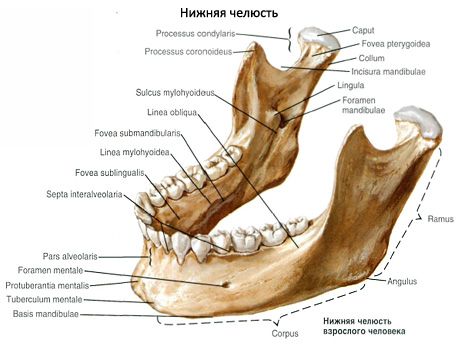
The lower jaw (mandibula) is the only movable bone of the skull. The unpaired lower jaw has a body and two branches.
The body of the lower jaw (corpus mandibulae) is curved forward with a convexity. The lower edge of the body, its base, is thickened and rounded, the upper edge forms the alveolar arch (arcus alveolaris). On the alveolar arch there are openings - dental alveoli (alveoli dentales) for 16 teeth, separated by thin bony interalveolar septa. On the outer surface of the alveolar arch there are convex alveolar elevations (juga alveolaria), corresponding to the alveoli. Along the midline in the anterior part of the body of the lower jaw there is a small mental protrusion (protuberantia mentalis). Behind and laterally from it at the level of the second premolar is the mental opening (foramen mentale).

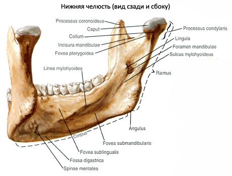
In the middle of the concave inner surface there is a small protrusion - the mental spine (spina mentalis). On the sides of it is the digastric fossa (fossa digastrica). Above the mental spine, closer to the alveoli, on each side is the sublingual fossa (fossa sublingualis) - a trace of the sublingual salivary gland. The mylohyoid line (linea mylochiodea) goes obliquely upward. Under it, at the level of the molars, is the submandibular fossa (fossa submandibularis) for the salivary gland of the same name.

The branch of the lower jaw (ramus mandibulae) is paired, goes upward and behind the body of the lower jaw. At the point where the body passes into the branch is the angle of the lower jaw (angulus mandibulae). On its outer surface is the masseteric tuberosity (tuberositas masseterica), and on the inner surface is the pterygoid tuberosity (tuberositas pterygoidea). On the inner surface of the branch of the lower jaw is the opening of the lower jaw (foramen mandibulae), leading to the canal of the same name, ending in the mental opening. From above, the branch of the lower jaw divides into two processes: the anterior coronoid and posterior condylar.

The coronoid process (processus coronoideus) is separated from the condylar process by the incisure of the mandible (incisure mandibulae). From the base of the coronoid process to the last molar runs the buccal ridge (crista buccinatoria).
The condylar process (processus condylaris) passes into the neck of the lower jaw (collum mandibulae), which ends in the head of the lower jaw (caput mandibulae).
What do need to examine?


 [
[