Medical expert of the article

New publications
Occipital bone
Last reviewed: 06.07.2025

All iLive content is medically reviewed or fact checked to ensure as much factual accuracy as possible.
We have strict sourcing guidelines and only link to reputable media sites, academic research institutions and, whenever possible, medically peer reviewed studies. Note that the numbers in parentheses ([1], [2], etc.) are clickable links to these studies.
If you feel that any of our content is inaccurate, out-of-date, or otherwise questionable, please select it and press Ctrl + Enter.
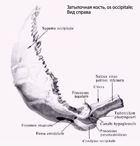
Occipital bone(os occipitale) is located in the posterior lower part of the cranial part of the skull. This bone is divided into the basilar part, two lateral parts and the occipital squama, which surround the large (occipital) opening (foramen magnum).
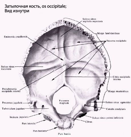
The basilar part (pars basilaris) is located in front of the large (occipital) opening. In front, it connects with the body of the sphenoid bone, together with which it forms a platform - a slope (clivus). On the lower surface of the basilar part there is an elevation - the pharyngeal tubercle (tuberculum pharyngeum), and along the lateral edge there is a groove of the inferior petrosal sinus (sulcus sinus petrosi inferioris).
The lateral part (pars lateralis) is paired and passes into the squama of the occipital bone at the back. Below each lateral part there is an elliptical elevation - the occipital condyle (condylus occipitalis), at the base of which is the hypoglossal nerve canal (canalis nervi hypoglossi). Behind the condyle there is a condylar fossa (fossa condylaris), and at its bottom is the opening of the condylar canal (canalis condylaris). To the side of the occipital condyle is the jugular notch (incisura jugularis), which, together with the jugular notch of the pyramid of the temporal bone, forms the jugular foramen. Next to the jugular notch on the cerebral surface is the groove of the sigmoid sinus (sulcus sinus sigmoidei).
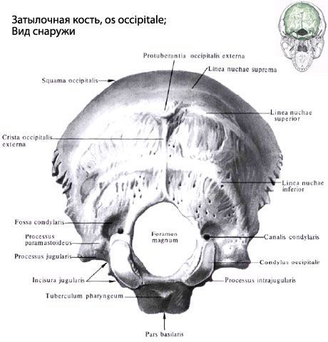
The occipital squama (squama occipitalis) is a wide, outwardly convex plate with strongly serrated edges. On the whole skull, they are connected to the parietal and temporal bones. In the center of the outer surface of the squama, the external occipital protuberance (protuberantia occipitalis externa) is visible, from which a weakly expressed superior occipital line (linea nuchae superior) extends in both directions. The external occipital crest (crista occipitalis externa) runs down from the protuberance to the large (occipital) opening. From its middle, the inferior occipital line (hinea nuchae inferior) runs to the right and left. The highest occipital line (linea nuchae suprema) is sometimes visible above the external occipital protuberance.
On the inner side of the occipital squama is a cruciform eminence (eminentia cruciformis), dividing the medullary surface of the squama into 4 pits. The center of the cruciform eminence forms the internal occipital protuberance (protuberantia occipitalis interna). To the right and left of this protuberance runs the groove of the transverse sinus (sulcus sinus transversus). Up from the protuberance runs the groove of the superior sagittal sinus (sulcus sinus sagittalis superioris), and down, to the large (occipital) opening, runs the internal occipital crest (crista occipitalis interna).
 [
[ Where does it hurt?
What do need to examine?
How to examine?