Medical expert of the article

New publications
Electroneuromyography
Last reviewed: 06.07.2025

All iLive content is medically reviewed or fact checked to ensure as much factual accuracy as possible.
We have strict sourcing guidelines and only link to reputable media sites, academic research institutions and, whenever possible, medically peer reviewed studies. Note that the numbers in parentheses ([1], [2], etc.) are clickable links to these studies.
If you feel that any of our content is inaccurate, out-of-date, or otherwise questionable, please select it and press Ctrl + Enter.
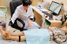
Electroneuromyography is performed for the purpose of topical diagnostics and assessment of damage to various parts of the peripheral neuromotor apparatus and determining the effectiveness of therapy for neuroinfections.
 [
[ Indications for electroneuromyography
- Development of motor deficit in an infectious disease associated (in the opinion of the attending physician) with damage to the peripheral nerves and/or muscles, early preclinical diagnosis of motor deficit.
- Evaluation of the effectiveness of therapy in a patient with neuroinfection with damage to the peripheral nervous system.
Preparation for an Electroneuromyography Study
Before the examination, the attending physician is warned about the need to stop prescribing drugs that affect neuromuscular transmission (proserin) 8-12 hours before the examination.
The study is conducted in the morning, before eating or 1.5-2 hours later. Before the electroneuromyography, the patient is reassured and informed about the procedure, the sensations he will experience, including the pain of electrical stimulation.
Electroneuromyography research technique
The examination is carried out in a supine or semi-recumbent position in a chair in a relaxed state.
Electroneuromyography uses two types of electrodes - superficial (cutaneous) and needle. Electromyographic recording of the action potentials of individual neuromuscular motor units is performed using needle electrodes. The evoked muscle potential (M-response) is recorded using superficial recording electrodes, which more objectively, compared to needle electrodes, reflect the total muscle activity. Atraumatic nature, no risk of infection, ease of use and relative painlessness of the study are the advantages of superficial electrodes. To find the locations of stimulating and recording electrodes, use the manuals and diagrams of J. A. DeLisa, K. Mackenzie, B. M. Gekht, L. O. Badalyan, I. A. Skvortsov.
When conducting electroneuromyography of the upper and lower extremities, a stimulating bipolar wick electrode and standard bipolar cutaneous recording electrodes are used. They are applied to the skin above the area of the muscle motor point: the main electrode is applied to the skin above the belly of the muscle being examined, and the indifferent electrode is applied to its tendon. Before applying the electrode, the skin is wiped with alcohol, and a special electrode gel is applied to the area of skin-electrode contact. The potential difference from the cutaneous electrodes is fed to the input of the electroneuromyography amplifier. A surface ground electrode is placed on the skin of the subject between the recording and stimulating electrodes. The felt wicks of the stimulating bipolar electrode are moistened with an isotonic sodium chloride solution before the study. The cathode of the stimulating electrode is placed above the motor point, and the anode is distal.
When conducting a comprehensive electrophysiological study, standard methods of stimulation electroneuromyography are used to determine the speed of impulse conduction along the motor fibers of peripheral nerves, terminal latency and the amplitude of muscle potential (M-response).
Contraindications to electroneuromyography
There are no contraindications to electroneuromyography (ENMG), but it is not recommended to use needle electrodes in patients with HIV infection, due to the high risk of infection of medical personnel during the study.
Interpretation of electroneuromyography results
Electroneuromyography reveals a decrease in the speed of impulse conduction along the nerves and a decrease in the amplitude of the nerve action potential not only with obvious clinical signs of mono- and polyneuropathy, but also in their absence. The decrease in the speed of impulse conduction detected in polyneuritis is used in the differential diagnosis of flaccid paralysis caused by acute neuroinfections ( poliomyelitis or polyneuritis).
Electroneuromyography can distinguish the nature of the damage to the peripheral nerves - demyelinating (characterized by a marked decrease in the speed of impulse conduction) or axonal (decrease in the amplitude of the M-response).
The extreme expression of pathology of the peripheral neuromotor apparatus is the absence of an M-response in electroneuromyography.