Medical expert of the article

New publications
Feeding disorder of the uterine myoma node
Last reviewed: 04.07.2025

All iLive content is medically reviewed or fact checked to ensure as much factual accuracy as possible.
We have strict sourcing guidelines and only link to reputable media sites, academic research institutions and, whenever possible, medically peer reviewed studies. Note that the numbers in parentheses ([1], [2], etc.) are clickable links to these studies.
If you feel that any of our content is inaccurate, out-of-date, or otherwise questionable, please select it and press Ctrl + Enter.
 [
[ Causes nutritional disorders of the uterine myoma node
According to modern concepts, uterine myoma is a dyshormonal tumor formed due to a disorder in the hypothalamus-pituitary gland-adrenal cortex-ovaries system. The dyshormonal nature of the tumor causes metabolic disorders, functional liver failure, and disorders of fat metabolism.
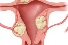
The tumor initially occurs intermuscularly, then, depending on the direction of growth, interstitial (in the thickness of the uterine wall), subserous (growing toward the abdominal cavity) and submucous (growing toward the mucous membrane of the uterus) tumor nodes develop. A capsule of muscle and connective tissue elements of the myometrium is formed around the myomatous node. In the presence of subserous nodes, the peritoneal cover of the uterus also participates in the formation of the tumor capsule; in submucous nodes, the capsule consists of a muscle layer and the mucous membrane of the uterus.
Most frequently (80%), there are multiple myomas of various sizes, shapes, and with different numbers of nodes. Single subserous or interstitial nodes are observed much less frequently. Subserous nodes are usually connected to the body of the uterus by a wide base, but sometimes they grow directly under the peritoneum, connecting to the uterus by a thin stalk. Such nodes are very mobile and are easily twisted. Submucous nodes are observed in approximately 10% of women with uterine myoma.
The frequency of uterine myoma necrosis, according to summary statistics, is about 7%. Tumor nodes are especially often necrotized during pregnancy, in the postpartum or post-abortion period.
Pathogenesis
Impaired blood supply in myomatous nodes is explained mainly by mechanical factors (torsion, bending, tumor compression). However, one cannot ignore the peculiarities of hemodynamics during pregnancy. Patients with uterine myoma during pregnancy experience a significant decrease in blood flow in the uterus, especially pronounced in the area of the intermuscular myomatous node, increased vascular tone, mainly in small-caliber vessels, severe difficulty in venous outflow, and decreased rate of blood filling of the arterial and venous bed. Clinical manifestations of changes in uterine hemodynamics are symptoms of increased myometrium tone, mild excitability of the uterus, and pain (pulling, aching, spastic).
Many authors have described various dystrophic processes in myomatous nodes (edema, necrosis foci, hemorrhage, hyaline degeneration, degeneration), which develop not only as a result of torsion of the pedicle of the subperitoneal node, but also as a result of ischemia, venous congestion, multiple thrombus formation in the intermuscular nodes of the tumor. A predisposing factor in this case is an increase in the size of myomatous nodes during the enlargement of the uterus during pregnancy.
There are dry and wet types of necrosis of uterine myoma. The so-called red necrosis of myoma has also been described. In dry necrosis, there is a gradual wrinkling of areas of necrotic tissue, forming peculiar cavernous cavities with remnants of dead tissue. In wet necrosis, there is softening and wet necrosis of tissue with subsequent formation of cystic cavities. Red necrosis is more common in intramurally located myomas. This form of necrosis usually occurs during pregnancy and the postpartum period. Macroscopically, the tumor nodes are colored red or brownish-red, have a soft consistency, and microscopically, pronounced varicose veins and their thrombosis are detected.
Some researchers see the cause of red necrosis in the increased tone of the myometrium surrounding the node with subsequent development of circulatory disorders in the tumor capsule and on the periphery. Necrotic changes are usually caused by circulatory disorders in the tumor. Aseptic necrosis is almost always accompanied by an infection that penetrates the node by hematogenous or lymphogenous routes. The causative agents of infection usually belong to the septic group of microbes (staphylococcus, streptococcus, E. coli). Infection of necrotically changed nodes of uterine myoma is very dangerous due to the real possibility of diffuse peritonitis and generalized infection (sepsis).
Symptoms nutritional disorders of the uterine myoma node
The leading symptom is pain in the lower abdomen of varying intensity depending on the type of nutritional disorder and the time of development of the process. Symptoms of general intoxication may also appear due to necrosis and infection of the tumor, tension of the anterior abdominal wall, possible increase in body temperature and leukocytosis.
Diagnostics nutritional disorders of the uterine myoma node
The diagnosis is based on the patient's complaints, which have a history of uterine myoma. The primary appeal of patients with a nutritional disorder of the myomatous node is possible.
During a vaginal examination, the presence of myomatous nodes in the uterus is determined, one of which is acutely painful upon palpation.
Ultrasound scanning facilitates the detection of difficult to palpate nodes and allows one to assess their condition.
A special role is played by the diagnosis of degenerative changes in myoma nodes in pregnant women, which often do not produce obvious clinical manifestations.
Of the instrumental methods, ultrasound of the uterus is of great importance in the diagnostic process, allowing to identify signs of a disruption in the nutrition of the tumor, as well as diagnostic laparoscopy, which makes it possible to visualize the node.
What do need to examine?
Who to contact?
Treatment nutritional disorders of the uterine myoma node
Patients diagnosed with myoma necrosis require urgent surgical treatment. Amputation or extirpation of the uterus is performed (most often, the fallopian tubes, which can serve as a source of infection, are removed at the same time). Conservative myomectomy is performed as an exception in young childless women under conditions of intensive antibacterial therapy in the postoperative period.
In some cases, conservative management of the patient and her preparation for a planned operation are acceptable. Such tactics are possible only when treating young women who do not have children. In order to improve the blood supply to the uterus, rheologically active agents (rheopolyglucin, trental) and antispasmodics (papaverine hydrochloride, no-shpa) are prescribed. If there is no quick effect from conservative therapy, surgery should be resorted to.
Treatment of impaired blood supply to uterine myoma nodes in pregnant women begins with conservative measures: antispasmodics, rheologically active drugs, tocolytics in combination with antibacterial and desensitizing agents are prescribed. If conservative therapy carried out for 2-3 days is ineffective, surgical treatment is indicated. Only subperitoneal nodes are subject to myomectomy. Impaired blood supply to intramural myomatous nodes requires removal of the uterus. In the postoperative period after enucleation of nodes, it is necessary to carry out treatment aimed at preserving pregnancy and preventing infectious complications.
Surgical (the scope of the operation is decided individually). In case of multiple uterine myoma in the perimenopausal period – amputation or extirpation of the uterus.
In case of secondary peritoneal phenomena and intoxication, it is also advisable to remove the uterus. In young women, organ-preserving surgery (myomectomy) is possible.