Medical expert of the article

New publications
Distal bite in children and adults
Last reviewed: 12.07.2025

All iLive content is medically reviewed or fact checked to ensure as much factual accuracy as possible.
We have strict sourcing guidelines and only link to reputable media sites, academic research institutions and, whenever possible, medically peer reviewed studies. Note that the numbers in parentheses ([1], [2], etc.) are clickable links to these studies.
If you feel that any of our content is inaccurate, out-of-date, or otherwise questionable, please select it and press Ctrl + Enter.
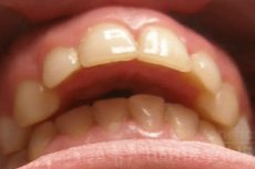
Incorrect positioning of the upper and lower jaws with a violation of the closure of the dental arches is a common orthodontic problem, and the most common type of pathological occlusion is considered to be distal bite (code K07.20 according to ICD-10).
Epidemiology
According to WHO statistics, the incidence of skeletal distal occlusion among Caucasian patients with occlusion problems is 38%, while among dark-skinned people it is no more than 20%. According to other data, the incidence of prognathic distal occlusion in the population does not exceed 26%.
Moreover, this type of bite disorder is observed in 80-85% of cases in childhood – during the period of eruption of baby teeth and their replacement by permanent ones. And only in 15-20% of cases does distal bite form in adults. [ 1 ]
Causes distal bite
Anatomical causes of malocclusion in the form of distal occlusion may be related to:
- with an increase in the size of the upper jaw - macrognathia (gnathos in Greek means jaw);
- with excessive development of the upper jaw (upper prognathism) and its forward protrusion, in which protrusion of the upper frontal teeth is observed;
- with mandibular micrognathia, hypoplasia, microgenia, or underdevelopment of the lower jaw (which in Latin is called mandibula);
- with the lower jaw recessed into the oral cavity and the upper jaw in the correct position - mandibular retrognathia;
- with simultaneous retrognathia of the lower jaw and prognathia of the upper jaw;
- with a posterior deviation of the dental arch of the lower jaw or a posterior position of its alveolar process - mandibular alveolar retrusion.
Many of the listed defects of the dental system are the result of improper formation of the visceral (facial) skeleton during the period of intrauterine development. In addition, congenital skeletal (jaw) distal and mesial bite (in which, on the contrary, the upper jaw is insufficiently developed, and the lower jaw is pushed forward) has a constitutionally inherited nature and can be observed in the family. [ 2 ], [ 3 ]
A deep distal bite in a child can be caused by:
- bilateral cleft palates - congenital non-fusion of the palate, as well as the alveolar process of the upper jaw and lip;
- congenital lower micrognathia, which occurs isolated in only 20% of cases, being a sign of a large number of syndromic disorders with varying degrees of developmental delay, in particular, Marfan, Seckel, Noonan, Apert, Crouzon, Pierre Robin syndromes, trisomy 13 ( Patau syndrome ), hemifacial microsomia, cri du chat syndrome, maxillofacial dysostosis ( Treacher Collins syndrome ), etc. [ 4 ], [ 5 ]
Also read:
Distal bite in adults can be formed due to maxillofacial injuries or pathological fractures of the jaws and/or their alveolar parts in the presence of a history of chronic osteomyelitis or fibrous ostitis, as well as due to degenerative changes in the temporomandibular joint (for example, with deforming osteoarthrosis).
Risk factors
The real and possible risk factors for the formation of distal bite include:
- heredity, that is, the presence of this orthodontic pathology in the family history;
- pathologies of pregnancy and various teratogenic effects on the fetus, increasing the likelihood of congenital defects of the facial skull;
- improper artificial feeding during infancy, prolonged use of a pacifier;
- dysphagia (swallowing disorders);
- childhood habit of sucking a finger, tongue or lip;
- anomaly of the tongue (glossoptosis) or shortening of its frenulum;
- incorrect eruption of milk teeth and disruption of its sequence;
- chronic enlargement of the tonsils and adenoids;
- habitual breathing through the mouth;
- changes in the dental arch – early loss of the first permanent molars or incisors;
- abnormal growth of permanent incisors;
- injuries to the facial bones, jaws and teeth;
- weakness of the chewing and orbicularis (circular) muscles of the mouth.
Pathogenesis
Orthodontists explain the pathogenesis of distal bite by genetic anomalies or congenital disproportions of the visceral skeleton, which manifest themselves in a forward shift of the upper jaw (prognathism) or a backward shift (retrognathism) of the lower jaw in such a way that the upper teeth are excessively protruded forward.
In addition, the mechanism of formation of mandibular prognathia-retrognathia in young children may be due to the above-mentioned physiological and functional factors. Thus, in infants, the lower jaw is initially shifted slightly backwards, and then - with the onset of the appearance of the first milk teeth - takes a normal position; bottle feeding does not provide the necessary load on the chewing muscles, and because of this, the lower jaw may remain insufficiently developed with the fixation of mandibular retrognathia. In this case, the situation is aggravated when this is a hereditary constitutional feature of the visceral skull. [ 6 ]
As for mouth breathing, it affects the position of the tongue in the oral cavity: it cannot perform a supporting function for the upper dental arch, and during the formation of the child’s dental system, this leads to lateral narrowing of the upper jaw, its prognathism and subsequent forward deviation of the upper incisors.
Symptoms distal bite
The following external and orthodontic symptoms of incorrect occlusion of teeth with distal occlusion are noted:
- anterior frontal displacement of the upper jaw;
- widening of the upper dental arch and shortening of the anterior part of the lower dental arch;
- backward displacement of the lower jaw or inward displacement (retrusion) of the lower incisors;
- overlap of the lower dental arch by the upper front teeth;
- an increase in the interocclusal gap between the upper and lower frontal teeth, which prevents the normal closure of the dental arches;
- pressure of the cutting edges of the lower incisors on the mucous membrane of the hard palate.
With a deep distal bite, the lower part of the face is shortened, and the upper row of teeth can almost completely obscure the lower row of teeth.
Obvious external signs of prognathic distal bite: the facial part of the skull is convex; the chin is beveled and shifted back; there may be a double chin; the lower labial and nasolabial folds are smoothed out, and the fold between the chin and lower lip is deep; the upper lip is shortened, and when smiling, the alveolar process of the upper jaw protrudes outward. Also, patients with upper prognathism may have gaps (tremas) between the crowns of the upper frontal teeth. [ 7 ]
And with a strongly protruding upper jaw, the patient’s mouth is constantly slightly open (due to the inability to close the lips), and the lower lip may be located behind the upper incisors.
Forms
The types or kinds of distal bite identified by specialists depend on the nature of the anomaly: it can be jaw, and in the case of an abnormal position of the upper jaw (prognathism) it is defined as a prognathic distal bite.
There is also a dental-alveolar type of distal occlusion: when there is an anterior protrusion of the maxillary dental arch and/or alveolar process (alveolar prognathism), or the upper incisors are tilted forward. The same type of occlusion is diagnosed when the mandibular dental arch or alveolar part of the lower jaw is tilted backward, or there is a deviation of the anterior lower teeth into the oral cavity.
In addition, there may be a combined bite - dental.
When the upper incisors overlap the crowns of the lower incisors by more than a third when the teeth are closed, a deep distal bite is defined. A distal open bite is characterized by the absence of closure of part of the upper and lower molars and the presence of a large vertical gap between their chewing surfaces. [ 8 ]
Complications and consequences
The main negative consequences and complications in the presence of distal occlusion and, especially, in cases of deep or open distal bite are:
- difficulty biting and chewing (and subsequent stomach problems due to insufficient chewing of solid foods);
- difficulty swallowing;
- functional disorder of the temporomandibular joint (with pain when opening the mouth and crunching when chewing);
- trauma to the soft palate by the lower incisors;
- hypertonicity of the masticatory muscle and bruxism;
- increased formation of tartar;
- increased wear and tear of the posterior molars and their deterioration;
- problems with articulation and diction.
Diagnostics distal bite
Diagnosis begins with a visual examination of the patient’s teeth and jaws, recording his complaints and collecting anamnesis.
By conducting teleradiography (or computer 3D cephalometry) and taking the appropriate measurements, the anatomical parameters of the facial skull and dental system are determined: the height of the face; the size of the nasolabial angle; the ratio of the position of the upper and lower jaws relative to the anterior part of the base of the skull; the angles of inclination of the alveolar processes of the jaws, the teeth themselves and their occlusal plane.
Instrumental diagnostics also includes:
- orthopantomogram – panoramic radiograph of the maxillofacial region;
- computed tomography or magnetic resonance imaging of the maxillofacial region;
- study of the tone of the jaw muscles (electromyography).
Differential diagnosis
Differential diagnostics based on cephalometric analysis data should clearly determine the type of malocclusion in order to select the optimal method of its correction.
Who to contact?
Treatment distal bite
To correct distal occlusion, there are various modifications of orthodontic structures and devices. First of all, with the dental-alvelar type of distal occlusion, braces are installed that correct the position of the teeth and dental arches in children (after the replacement of milk teeth with permanent ones), adolescents and adults.
Additionally, in bracket systems that exert pressure on the dental arch, an individually manufactured multi-loop arch is used for distal skeletal bite. With its help, it is possible to correct defects of the dental arch, often accompanying prognathism. The brackets and the loop are worn constantly and for a long time, and after their removal - to consolidate the results of the correction - removable or stationary retaining devices are placed on the inner surface of the teeth for some time: orthodontic retention plates or orthodontic splints (retainers).
And to change the abnormal tilt of the front teeth of the upper row and stimulate the orbicularis muscle, the installation of vestibular plates in children is practiced.
Instead of plates, a trainer for distal occlusion of the dental-alvelar type is sometimes used, which is a silicone alignment brace-trainer, put on the teeth for their correct positioning. Before orthodontic treatment (since the installation of braces is carried out only on permanent teeth), children with problems with occlusion, from the age of six (with the beginning of the period of mixed occlusion), can install a pre-orthodontic type trainer. [ 9 ]
In some cases of distal occlusion of jaw origin during the period of visceral skull growth, it is possible to treat distal occlusion without surgery. For this purpose, functional orthodontic devices for distal occlusion can be used:
- bionators (Balters and Janson), consisting of plates and arches, the adjustable force action of which contributes to the increase of the body and branch of the lower jaw and its anterior displacement;
- Frenkel functional regulator (two modifications), used to correct this occlusion disorder during the active growth of children at the end of the period of eruption of baby teeth and at the beginning of their replacement with permanent teeth;
- Herbst and Katz appliances with support on teeth, stimulating the growth of the lower jaw by correcting the contraction of the orofacial muscles;
- Forsus stationary device for the upper and lower dental arches, which allows for the simultaneous retraction of protruding upper incisors back and the pulling of lower teeth forward in adolescent patients;
- a semi-rigid corrective device TwinForce fixed to both dental arches for deep distal bite with mandibular retrognathia. Similarly, the use of the Twin Block device is TwinBlock for distal bite with mandibular hypoplasia; the structure is attached to the dental arches in such a way that the anterior position of the lower jaw is ensured and the occlusal relationships of the dental arches are normalized. [ 10 ]
Can aligners or veneers correct a distal bite? Clear aligners, made from a mold of the patient's jaw, are essentially modernized mouth guards, and they can fix the dentition without affecting the alveolar process of the upper jaw. Therefore, these dental onlays (they are worn 24 hours a day, removed before eating) can help reduce the anterior inclination of the upper incisors. [ 11 ]
But veneers that improve the appearance of the front teeth are not installed on the distal bite: this is an aesthetic dentistry procedure that cannot straighten an abnormally positioned row of teeth. Their installation can only be performed after orthodontic treatment, for example, to change the shape of the crowns of the front teeth in the presence of large interdental spaces.
Surgical treatment, operations
According to foreign clinical statistics, surgical treatment of distal occlusion is performed in approximately 5% of patients with a skeletal type of prognathic bite with pronounced maxillofacial defects, ankylosis and degenerative changes in the temporomandibular joint. [ 12 ]
Orthognathic surgery involves performing an operation for distal occlusion, which is aimed at correcting pathological changes in the dental system - prognathia or micrognathia, which are rarely treatable with braces, plates and other devices for correcting occlusion.
Maxillofacial surgeries are performed for cleft lip and palate, osteotomy of the upper jaw - with retrotransposition (movement backwards) of its frontal part and fixation in the desired position (with permanent titanium fasteners). In adult patients with open distal bite, compact osteotomy can be performed.
In the presence of mandibular retrognathia, various techniques of osteotomy of the lower jaw can be used. [ 13 ]
Exercises for distal bite
For normal functioning of the orofacial muscles and temporomandibular joints, it is recommended to do exercises for distal bite and other disorders of the dental system. Exercises for the masticatory, pterygoid, orbicularis and other maxillofacial muscles are related to myofunctional therapy, which helps to increase the effectiveness of the use of orthodontic devices. [ 14 ]
Special myogymnastics for distal bite should be done daily - twice for five to ten minutes. Here are some of the basic exercises:
- wide opening and closing of the mouth (several repetitions);
- maximum possible forward extension of the lower jaw;
- puffing out your cheeks vigorously, holding the air for 10 seconds and slowly blowing it out (this exercise can be done with water);
- pursing the lips and then stretching them (as if smiling);
- retraction of the tongue to the base of the palate (with the mouth closed).
Prevention
In case of hereditary features of the anatomy of the visceral skull and in children with syndromic anomalies of the jaws, which are congenital and genetically determined, prevention of distal bite is impossible.
Experts believe that the main preventive factors for the development of distal bite in a child are natural breastfeeding (and if artificial, then properly organized), refusal of a pacifier, weaning off the above-mentioned habits, etc. It is necessary to promptly treat everything that can prevent the child from breathing freely through the nose.
Forecast
With the dental-alveolar type of distal occlusion, the prognosis regarding the results of hardware orthodontics is much better than with the jaw type, when it is necessary to resort to orthognathic surgery.
In adults, correcting defects of the dental system is very difficult, time-consuming and expensive, and predicting the outcome of their correction is even more difficult.