Medical expert of the article

New publications
Diffuse changes of myometrium by type of adenomyosis, endometriosis, focal, nodular: what does it mean?
Last reviewed: 12.07.2025

All iLive content is medically reviewed or fact checked to ensure as much factual accuracy as possible.
We have strict sourcing guidelines and only link to reputable media sites, academic research institutions and, whenever possible, medically peer reviewed studies. Note that the numbers in parentheses ([1], [2], etc.) are clickable links to these studies.
If you feel that any of our content is inaccurate, out-of-date, or otherwise questionable, please select it and press Ctrl + Enter.
Such a conclusion of a gynecologist as diffuse changes in the myometrium does not bode well for a young woman, because such a violation of women's health often threatens that she will not be able to become a mother of her own child. When diffuse changes appear, a woman is most often diagnosed with "endometriosis", and this means that her chances of getting pregnant and carrying a child are extremely small. So what kind of disease is this that prevents the most noble and beautiful dream of every woman from coming true - the opportunity to give life to a new person?
What is myometrium?
Before we begin to consider the issue of diffuse and other changes in the tissues of a woman's body, we must first understand what tissues we are talking about. One of the main organs of the female reproductive system is the uterus. It is in it that during the first 7-9 months after conception a little person is formed, grows and develops - a miniature copy of its father and mother. It is thanks to the rhythmic contractions of the walls of the uterus that a son or daughter at the right time gets the opportunity to go beyond the mother's body and see the world.
The uterus in a woman is a pear-shaped organ located in the center of the pelvis. Its closest neighbor on one side is the urinary bladder, and on the other side is the rectum. Depending on how full they are, they can tilt the uterus forward or backward a little.
The uterus is considered a hollow organ, in which there is only empty space for the time being. The organ itself consists of three sections: the fundus, the body and the cervix, which flows into the vaginal cavity.
The walls of the uterus also have 3 layers:
- the outer or serous layer, identical with the lining of the bladder and considered to be its continuation, is called the perimetrium,
- the inner or muscular layer, which is the thickest and is a collection of muscle and elastic fibers, as well as connective tissue, is called the myometrium,
- The inner layer or mucous membrane, which consists of a basal and functional layer and is a layer of columnar epithelium attached to a connective tissue base, is called the endometrium.
The myometrium, the diffuse changes of which we have undertaken to consider in this article, in turn is a multilayered tissue:
- the outer or subserous layer is a thin tissue of longitudinal and some circular fibers, tightly attached to the perimeter,
- the middle or vascular layer is the strongest and thickest part of the myometrium, consisting of circular fibers and abundantly supplied with blood vessels,
- The inner or submucosal layer is again a thin tissue, which is represented by longitudinal fibers and is tightly adjacent to the endometrium.
When we talked about the fact that the uterus not only preserves the human fetus inside itself during pregnancy, but also helps it to come out when the baby's body is already capable of independent existence. Unfortunately, sometimes, due to certain disorders, the fetus has to leave the mother's womb ahead of time, when it is not yet viable, and in such a case we talk about a miscarriage or premature birth.
How does the uterus help push the baby out into the world? With the help of its inner layer – the myometrium. Rhythmically contracting, it helps the baby move through the birth canal. It is clear that the condition of this layer largely determines whether a woman can carry a pregnancy and give birth to a child on her own. And any changes in the muscular layer of the uterus cannot but affect its functionality.
Normally, the myometrium lines the walls of the uterus with a uniform layer, i.e. its thickness is approximately the same and no pathological compactions or voids are found inside the muscular layer. Such a muscular layer functions normally. We feel its noticeable contractions during menstruation, as well as before and during childbirth.
But the endometrium changes its thickness significantly during the menstrual cycle: from 1-2 mm at the beginning of the cycle and up to 15 mm during menstrual bleeding. Both of these layers are closely related to each other, so pathological changes in the endometrium often affect the inner layer of the uterus, disrupting its functionality.
Diffuse changes in the myometrium are diffuse disturbances in the structure and functionality of the inner layer of the uterus, which affect the entire organ, not just its individual parts. And the severity of such changes determines a woman's health and ability to become a mother.
Epidemiology
According to statistics, endometriosis is one of the most common pathologies of the female reproductive system (although there are cases of this disease in men, affecting the pelvic organs). The number of women with such a diagnosis is steadily approaching the number of patients with inflammatory pathologies of the pelvic organs.
In practice, it has been noted that half of the cases of adenomyosis (proliferation of epithelial cells inside the uterus) and endometriosis (germination of endometrial cells into other nearby organs) are combined with thyroid diseases (most often we are talking about an autoimmune pathology called "thyroiditis", characterized by a chronic inflammatory process in the tissues of the thyroid gland, or dysfunction of the pituitary gland). This allows us to suspect these pathologies of involvement in the development of dysplastic processes in the uterus and adjacent tissues.
The percentage of women of reproductive age suffering from endometriosis to varying degrees worldwide is approaching 10-11%. Severe and moderate diffuse changes in the myometrium, which indicate the development of endometriosis, are detected in more than 30% of women who have been diagnosed with infertility. About 75% of women with dysplastic changes in the uterine tissues cannot have children.
Causes diffuse changes in the uterine myometrium
If a woman sees the words "heterogeneous myometrium" in the ultrasound results, she, of course, begins to worry about what this could mean for her. The very concept of heterogeneity of the inner layer of the uterus speaks of diffuse changes in it. But these changes need to be considered in an age context.
In the postmenopausal period, a non-uniform myometrium is considered a normal variant. Changes in a woman's hormonal background at this time dictate their own rules. In reproductive age, during menstruation, the mucous layer of the uterus thickens, and when menopause comes, such changes are no longer observed. The endometrium becomes thinner, and since it is directly connected to the myometrium, degenerative processes affect it as well.
For women over 45 years of age after menopause, diffuse changes in the myometrium do not pose a danger. This is a natural physiological process of aging caused by hormonal changes. Pregnancy and the desire to have a child at this age are usually no longer a concern, so myometrium heterogeneity in ultrasound results can simply be ignored unless there is a suspicion of oncological processes.
But at a young age, when most women dream of becoming a mother, changes in the structure and functionality of the strongest muscular layer of the uterus pose a real threat to a woman's dream and her health. Normally, the uterine endometrium is tightly adjacent to the myometrium. If the cells of the mucous layer begin to penetrate into the muscular layer, they speak of the initial stage of endometriosis - adenomyosis. Deeper germination of the endometrium into the myometrium and perimetrium is called endometriosis. When the process goes beyond the uterus, doctors diagnose "ectopic endometriosis".
If the endometrium does not grow into the muscular layer, and its thickness increases only due to the growth of cells into the uterine cavity, doctors talk about the borderline diagnosis of "dysplasia of the uterine endometrium" (more often dysplasia of the cervix, if the process affects not the entire organ, but only its final rounded part).
The mechanism of diffuse changes in the myometrium and endometrium has not yet been fully studied. Doctors have several theories of the development of the pathological process. Some specialists are looking for reasons for changes in the tissues of the uterine wall in genetic predisposition, but they directly associate the formation of the lesion with hormonal changes in the body. Scientists believe that against the background of a violation of hormone production, the process initially embedded at the DNA level is activated and the endometrial cells begin to grow uncontrollably both inside and outside the uterus, damaging the structure of the muscle layer.
The hormonal theory is supported by changes in the thickness of the endometrium in different phases of the menstrual cycle, caused by changes in the hormonal background. This theory is also supported by the fact that during pregnancy and menopause the process is in the opposite direction, i.e. the thickness of the endometrium becomes smaller, as at the beginning of the menstrual cycle.
The second leading theory of the development of dysplasia and endometriosis is considered to be the implantation theory. According to it, the pathogenesis of the disease is based on the ability of rejected endometrial cells to form foci of diffuse changes in the form of tumor processes under certain unfavorable conditions.
Risk factors
According to this theory, the risk factors for the appearance of diffuse and focal changes in the myometrium are:
- previous abortions and curettages, which damage the inner layer of the uterus (and the more frequent the abortions, the higher the risk of developing a pathological process),
- any other interventions in the uterus, including cesarean section, curettage in case of severe inflammatory processes, removal of cystic formations and polyps, surgical treatment of uterine fibroids, which are benign tumors, surgical treatment of oncological diseases in the uterus,
- infection of the uterine tissue (infection entering the uterine cavity, which often happens during menstrual bleeding, when the cervix is slightly open, causes an inflammatory process in the endometrium, which then provokes dysplastic changes in the inner, and then the middle layer).
Now, regarding the unfavorable conditions that increase the risk of endometriosis. These are:
- inflammatory and especially infectious-inflammatory diseases of the internal and external genital organs, pelvic organs (risk factors include promiscuous sexual relations and poor intimate hygiene),
- hormonal imbalance and stressful situations that cause disruptions in the neuroendocrine system,
- any endocrine diseases that cause hormonal and metabolic disorders,
- dysfunction of the endocrine glands (in addition to the ciliated columnar epithelium, the endometrium contains secretory cells),
- any tumor processes in the uterine cavity,
- anemia, immune system disorders, decreased body defenses due to existing chronic diseases,
- nutritional disorders with deficiency of vitamins and microelements,
- bad habits: smoking, alcohol abuse, addiction to drinks containing caffeine, as well as uncontrolled intake of medications,
- complicated pregnancy and childbirth,
- negative impact of solar UV radiation (if there is a predisposition to endometrial dysplasia, prolonged or frequent exposure of the body to sunlight can provoke the development of the pathological process),
- the release of blood with particles of epithelial cells during menstruation into the fallopian tubes and their deposition on the ovaries can provoke diffuse changes in the myometrium and ovaries.
Depending on where exactly the rejected endometrial cells end up with menstrual blood and where they begin to actively divide, diffuse changes can cover various parts of the female reproductive system and beyond. If the proliferation of endometrial cells is observed not only inside the uterus itself, but also in its terminal section, which connects to the vagina, we speak of diffuse changes in the myometrium of the body and cervix.
The entry of such cells into the vagina, bladder and peritoneum with their settling on the walls of organs under appropriate conditions can cause endometriosis of the vagina, bladder or peritoneum. If the endometrial cells grow into the tissues of the rectum, this is called the rectovaginal form of endometriosis.
There is another hypothesis, called metaplastic. According to this version of the development of events, rejected endometrial cells do not take root on the walls of the uterus and other organs near it, but provoke metaplastic changes in other cells. This hypothesis is supported by the fact that in some cases endometritis can degenerate into malignant tumors.
 [ 7 ]
[ 7 ]
Symptoms diffuse changes in the uterine myometrium
As we can see, the outlook for young women with frequent abortions and cleanings is not very pleasant. At some point, they may hear a frightening diagnosis, without even suspecting that something is wrong with their reproductive system. The fact is that diffuse changes in the myometrium may not make themselves known for a long time, because until a certain time they are not considered a pathology.
We have already mentioned that such changes in the uterine tissues are considered normal during menopause, and some healthy young women have a heterogeneous structure of the myometrium, which is inherited. In the latter case, we are talking about a weakly expressed heterogeneity, in which the difference between the layers is insignificant, and other parameters of the uterus (the size of the organ and the thickness of its walls) are within the normal range. Women usually do not feel such changes in the uterine tissues at all.
But pathological changes caused by provoking factors can make themselves known. However, a woman may simply not pay attention to such non-specific symptoms or attribute their appearance to other causes. So the first signs of endometriosis can be:
- mild pulling or squeezing pain in the lower abdomen, which most often appears during ovulation, as well as on the eve of menstruation (these symptoms are often completely ignored by women),
- quite severe pain during menstruation (the cause may also be a low pain threshold, endometritis, congenital anomalies in the structure of the uterus),
- pain during intercourse, which many attribute to the discrepancy between the sizes of the male and female genitals, the inexperience of the sexual partner, the incorrect position of the uterus (its bending),
- vague pain during urination and defecation,
- too heavy menstrual bleeding,
- menstrual cycle disorders (this symptom is characteristic not only of pathologies of the genitourinary system),
Later, spotting and slight bleeding between periods appear, which make the young woman wary, especially if they are repeated several times.
Mild pain in the lower abdomen as endometriosis of the uterus progresses can become longer and more intense, radiating to the groin and lower back. Often, the appearance of chronic pelvic pain is the reason for visiting a doctor, although with regular gynecological examinations, changes in the size and condition of the uterus can be detected much earlier.
Stages
According to doctors themselves, diffuse changes in the myometrium are not considered a full-fledged diagnosis. By the term heterogeneous myometrium, they mean a deviation from the norm, which does not necessarily have to be considered a pathology. Minor changes in the structure of the endometrium and myometrium can equally turn out to be a congenital feature or an acquired pathology.
But moderate diffuse changes in the myometrium already indicate the initial stages of a serious pathology - endometriosis. Since endometriosis originates in the uterus, then in the early stages of the disease, when only the tissues of the organ itself grow, we should rather talk about adenomyosis.
Diffuse changes in the myometrium by the type of adenomyosis are tissue damage inside the uterus. At the first stage of this disease, some changes in the endometrium are noted, and its individual cells can be found in the submucosal layer of the myometrium. The second stage is already characterized by damage to almost half of the muscle layer, while at the third stage, penetration of epithelial cells to a depth of more than half the thickness of the myometrium is noted. The fourth stage of the disease is damage to the entire muscle layer and organs close in location, i.e. endometriosis itself.
If the cause of the change in the structure of the various layers of the uterine wall was mechanical damage during medical and diagnostic procedures and abortions, ultrasound may show diffuse focal changes in the myometrium. This indicates that not the entire myometrium lining the uterus is subject to change, but only individual areas of various localizations, ranging in size from 2 mm to 1.5 cm (areas where the uterine wall was damaged).
It is also possible that multiple individual small foci (up to 5-6 mm) of a round shape without clear contours and a superficial capsule may appear. In this case, they speak of diffuse nodular changes in the myometrium.
Complications and consequences
Diffuse changes in the myometrium are themselves common disorders of the structure of the uterine wall, when the elasticity and functionality of the muscular layer are disrupted by the introduction of looser endometrial cells into it. For a woman who does not plan to become a mother, such a situation seems quite safe, especially if there is no discomfort or unpleasant sensations. But the process can gradually cover an increasingly large area of the uterus and move on to other organs, so leaving such a disorder untreated means incurring new health problems.
Gradually, diffuse changes in the uterine wall will develop into adenomyosis, or even spread beyond the uterus. This will be accompanied by the appearance of various symptoms of the disease (usually symptoms appear at stage 2 or 3 of the disease). Painful periods and heavy blood loss quite often lead to the development of iron deficiency anemia. In addition to weakness, dizziness, increased fatigue, shortness of breath and fainting, this disease entails a decrease in the body's resistance to infections. Moreover, such patients are more susceptible to stress factors, so they are more often diagnosed with neuroses.
Pain during menstruation is compounded by pain during sexual intercourse, which prevents a woman from getting the desired pleasure. Dissatisfaction with sex leads to increased irritability and conflict. Regular refusals of a woman to have sexual intercourse often become the cause of discord in the family.
Many young women are concerned about the logical question: is it possible to get pregnant with diffuse changes in the myometrium? It is impossible to answer this question unambiguously. Although there is a lot of information that women with such a diagnosis experience great difficulties not only with carrying a pregnancy, but even with conceiving a child. With the development of adenomyosis, in half of the cases, there is a violation of the structure of the endometrium and the development of an adhesion process that prevents the fertilized egg from entering the uterus (often this situation ends in an ectopic pregnancy).
But even if conception has occurred and the egg has been implanted in the uterus, there is no guarantee that it will stay there for 9 months and the baby will be born on time. Diffuse changes in the myometrium during pregnancy are the main risk factor for miscarriages and premature births. Dysplastic changes in the uterine tissues are accompanied by intermenstrual bleeding, which in turn provokes inflammatory processes. Inflammation of the uterine tissues and increased tone poses a threat of early termination of pregnancy.
As for older women and those who seem to be in no danger, there is no need to relax here either. The inflammatory process in the area of the regularly bleeding uterus leads to the formation of adhesions, causing chronic nagging pain and fusion of the pelvic organs. If endometrial cells extend beyond the uterus and begin to grow on the walls of other organs, they also begin to bleed. Against this background, cysts can form in the ovaries, the cavity of which is filled with menstrual blood.
The growth of uterine tissue leads to its enlargement. The uterus compresses other nearby organs and can cause neurological pain.
But the greatest danger of such complications as anedomiosis and endometriosis is considered to be the transformation of tumor cells into malignant ones. And although the risk of such a transformation is small (no more than 3%), its consequences are so terrible that it is impossible not to take this possibility into account.
Diagnostics diffuse changes in the uterine myometrium
Since diffuse changes in the myometrium are considered a pathological condition that may not give any symptoms at first, they are usually detected during a routine examination or during an ultrasound examination (for example, during pregnancy or the inability to become pregnant for a long time). It is clear that a gynecologist cannot see such changes visually during an examination on the chair, but the growth of uterine tissue is accompanied by its enlargement and change in shape (it takes on the shape of a ball), which is what the specialist will detect.
A gynecological examination, which is best done the day before menstruation, may show the presence of tubercles and nodes on the surface of the organ and in nearby tissues. If such a picture is also confirmed by the patient's complaints of painful and heavy periods that last 6-7 days, pain during sexual intercourse, the appearance of symptoms of anemia, the doctor can make a preliminary diagnosis - adenomyosis. If there are no complaints, a borderline condition is suspected, which can develop into a disease or remain at the same stage.
To assess the condition of vital organs and prescribe treatment, a woman is prescribed a standard set of laboratory tests: general urine analysis and clinical blood test. These same tests will help to identify and assess the degree of the inflammatory process in the woman's body (without determining its localization) and the presence of malignant cells. To assess the state of the hormonal background, which has a direct impact on the development of diseases with diffuse changes in the myometrium and endometrium, a blood test for hormones is prescribed.
A vaginal smear is also mandatory; microscopy of it will not only reveal the presence of infections (bacteria, viruses, fungi, etc.) in the reproductive system, but will also allow the detection of a large amount of cylindrical epithelium secreted from the uterus during inflammatory and dysplastic processes.
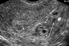
And yet, although tests complement the existing picture, they do not by themselves allow confirming the diagnosis. But instrumental diagnostics succeeds in this. Ultrasound diagnostics comes to the fore here. If diffuse changes in the myometrium are suspected, the patient is prescribed an ultrasound of the uterus or pelvis. On the computer screen, the doctor can not only see changes in the size of the uterus, but also measure the thickness of its walls, carefully examine pathological foci.
Echographic signs of diffuse changes in the myometrium allow the doctor not only to make a diagnosis with 90% reliability, but also to assess the degree of development of the disease. Different tissues of the body have different ability to reflect ultrasound waves, so echogenicity is an important criterion for ultrasound diagnostics. Increased echogenicity of a tissue area indicates the presence of diffuse changes in it. Blurred contours and heterogeneity of the myometrium also indicate such changes.
Hyperechogenic areas in the uterus indicate compactions in its tissues. In the diffuse form of adenomyosis (endometriosis), small compactions are noted over the entire surface of the uterus, i.e. the myometrium has a cellular structure. Hyperechogenic inclusions are scattered over the entire area of the organ and have limited dimensions (up to 5 mm).
The size of the uterus plays a major role in diagnosing the pathology. So, in women who have not given birth, the cervix can be 2-2.5 cm wide, and the length and thickness will be within 2.5-3.5 cm. The body of the uterus: length and thickness within 3.8-5 cm, width 2.7-3.7 cm. Pregnancy and childbirth have little effect on the size of the organ, however, as do age-related changes in the menopause period.
However, a normal noticeable increase in the uterus can only be noted during pregnancy as the fetus grows and develops inside it. During the first 2 months of pregnancy, the uterus increases in size by 3 times. With endometriosis, the size of the uterus will be approximately the same as that of an expectant mother whose pregnancy period is from 5 to 9 weeks. It turns out that the uterus will increase in size by 1.5-3 times.
At the initial stage of endometriosis, ultrasound may not show significant changes in the myometrium. Small hyperechoic inclusions may even remain unmarked. BUT the greater the thickness of the endometrium, the more clearly the echo signs of diffuse changes appear.
Diffuse changes in the myometrium such as adenomyosis at any stage of the pathology can be established using an endoscopic examination - laparoscopy. The study also allows you to assess the degree of tissue proliferation and even take material for histological examination for the presence of malignant cells. With the help of laparoscopic equipment, you can also carry out therapeutic manipulations, for example, cauterize pathological foci.Hysteroscopy has similar capabilities.
[ 19 ]
Differential diagnosis
Differential diagnostics, which consists of comparing the results of various studies and the patient's medical history, allows us to differentiate a congenital anomaly of the myometrium structure from:
- diffuse changes in the muscular layer observed in adenomyosis, endometriosis, endometritis (inflammation of the uterine mucosa),
- cervical cysts,
- diffuse form of chronic metritis, which is an inflammation of the inner and middle layers of the uterine wall,
- endometrial hyperplasia,
- uterine polyposis,
- proliferation of the follicular apparatus,
- oncological diseases of the reproductive system.
If there are certain difficulties in making a diagnosis, they resort to MRI. This study provides 99% accuracy of diagnosis.
Treatment diffuse changes in the uterine myometrium
Read more about traditional treatment of diffuse myometrial changes in this article.Folk remedies and herbs are also used.
Prevention
As it usually happens, we usually turn to our unloved doctor when we start to be bothered by pain in the lower abdomen, incomprehensible spotting, regular pain during intercourse. As long as nothing bothers a woman, she is in no hurry to go to the doctor. Unless she is forced to do so by a delay in menstruation, indicating the onset of pregnancy or menopause.
But diffuse changes in the uterine tissues occur gradually and progress gradually. A woman may not suspect such disorders for years until unusual alarming symptoms appear. Regular visits to a gynecologist 1-2 times a year would help to identify these changes much earlier in order to take measures to prevent the spread of the pathological process and preserve the reproductive function of a young woman.
But prevention of diffuse and focal changes in myometrium tissues is not only considered to be regular visits to the gynecologist. Most of the fair sex strive to look even more beautiful, showing off an even bronze tan, which can be obtained in a solarium or on the seaside. But does everyone know what the price of this tan is?
The negative impact of ultraviolet rays affects not only the skin, where melanoma foci may subsequently be found, but also internal tissues. Under the influence of active UV radiation, hyperplastic processes in the female reproductive system are activated. And a tanning lover may soon be diagnosed with infertility caused by adenomyosis or endometriosis.
It is important to understand that a beautiful, even tan can be obtained at a lower cost by relaxing at sea during the "velvet season" or basking in the inactive spring sun rays. In the summer, it is recommended to sunbathe only in the morning and evening. Of course, such a tan will not be noticeable so quickly, but its consequences will not be so sad.
Since hyperplastic pathologies are associated with a woman's hormonal imbalance, it is worth taking care of it. If hormonal imbalances are not hereditary, then they arose due to certain life situations: stress and anxiety, decreased immunity, metabolic disorders, infectious diseases, parasites. If you treat such diseases in time, maintain your immunity at a high level and take care of the health of your nervous system (learn how to react correctly to stressful situations), hormonal imbalances can be prevented, which means you can avoid disorders in the reproductive organs (and not only!).
Prevention of all kinds of pathologies caused by hormonal disorders will be maintaining a normal weight, proper nutrition, an adequate attitude to taking medications, adequate sleep, and giving up bad habits.
If it is still not possible to avoid the development of the pathological process, it is important to begin treatment of the disease detected during the gynecological examination as soon as possible. Since adenomyosis and endometriosis are prone to frequent relapses, the woman will subsequently have to continue taking oral contraceptives or insert a hormonal intrauterine device for preventive purposes. The device will protect her not only from pregnancy, but also from hyperplastic processes in the uterus for 5 years.
For women who would like to become a mother, there is also good news. It turns out that pregnancy significantly reduces the risk of endometrial tissue growth and its transfer to the walls of other organs, because during this period there is no menstruation and the woman's hormonal background is somewhat changed, which does not contribute to hyperplastic processes. It can be said that pregnancy is a preventive measure against endometriosis.
But this cannot be said about abortions and other surgical interventions on the uterus. Women planning to kill an unborn child should think a thousand times about the consequences of a curettage or premature birth, whether they will be able to get pregnant again and create a full-fledged family after this.
Did the reader find the prevention of pathological processes in the female reproductive system too complicated, since it requires giving up the usual way of life? Probably, if we discuss the prognosis of diseases developing against the background of diffuse changes in the tissues of the uterus, the view on prevention with all its complexities may change significantly.
We will not talk about the difficulties that a young woman will have to endure if she ignores the problem or refuses treatment. We have already discussed this issue in the section "Consequences and complications". But what can threaten a woman who has undergone appropriate treatment while preserving the uterus and its functions?
It should be noted that about 20 percent of women visit a gynecologist again with the same problem during the first year after completing a course of hormonal therapy or gentle removal of hyperplasia foci. Over the next 4 years, 70-75% of women visit a doctor, and only 25% can forget about the disease for a long time, reaching the onset of menopause.
The best prognosis is observed after removal of the uterus, but in this case the woman considers herself inferior and will never be able to have her own children (unless they are carried by a surrogate mother, and only if the ovarian function is preserved). It turns out that any methods of treating endometriosis and adenomyosis do not guarantee a complete cure with the ability to have children preserved. Isn't this a serious reason to take a different look at the difficulties of disease prevention?
Diffuse changes in the myometrium are not a death sentence, but there is no longer talk of normal functioning of the uterus. The sooner the pathological process is detected, the greater the chances of emerging victorious and preserving the function intended for a woman by nature itself, experiencing the joy of motherhood and protecting yourself from pain and suffering, both physical and psychological. Understanding this comes gradually, and the sooner a woman realizes the need to keep her body healthy, the greater her chances of becoming a happy mother, and perhaps more than once.
Forecast
Diffuse changes in the myometrium, if they are not a hereditary feature, are considered a pathological process. And although in the initial stage they are not yet considered a disease, they may well lead to it in the case of progression of the hyperplastic process.
Adenomyosis, endometriosis, uterine myoma and some other disorders of women's health are the consequences of untimely detection of pathological changes in the tissues of the uterus. And this means that the woman turned to the gynecologist too late.