Medical expert of the article
New publications
Diagnosis in dermatology using the Wood's lamp
Last reviewed: 29.06.2025

All iLive content is medically reviewed or fact checked to ensure as much factual accuracy as possible.
We have strict sourcing guidelines and only link to reputable media sites, academic research institutions and, whenever possible, medically peer reviewed studies. Note that the numbers in parentheses ([1], [2], etc.) are clickable links to these studies.
If you feel that any of our content is inaccurate, out-of-date, or otherwise questionable, please select it and press Ctrl + Enter.

For almost a century, a simple, safe and quite effective method of detecting certain skin infections and pigmentary disorders has been the diagnosis in dermatology using a Wood's lamp, which projects long-wavelength ultraviolet light onto the skin.
What's a Wood's lamp?
American optical physicist Robert Williams Wood (1868-1955) was a pioneer in infrared and ultraviolet photography, and in 1903 it was for UV photography that he developed the UV filter, which transmits ultraviolet radiation in the 320-400 nm wavelength range and blocks most visible light. That is, it is the long-wavelength rays of the UV-A spectrum that predominate in sunlight and penetrate deeper into the skin; they are invisible, which is why they are called black light. UV-A black light allows the naked eye to observe fluorescence, the colored glow that many substances emit when exposed to it. [1]
Based on this filter (Wood's glass), the scientist created the blacklight lamp, which after World War I found application in some fields, particularly in forensics. Later, the Wood's lamp found application in other scientific fields, including emergency medicine, ophthalmology, [2] gynecology and veterinary medicine. [3], [4], [5] The Wood's lamp was used in dermatology in the mid-1920s to diagnose a number of fungal skin diseases (mycoses), bacterial infections and pigmentation abnormalities.
Healthy normal skin under the Wood's lamp looks blue and does not glow, but areas of thickening of the epidermis give a white glow, areas of increased oiliness of the skin can be seen in the form of yellow spots, and dehydrated areas have the appearance of purple spots.
But some fungi (dermatophytes), bacteria or changes in the pigmentation of the patient's skin when exposed to UV-A rays can cause discoloration of the affected area.
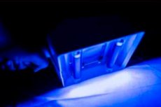
What does a Wood's lamp look like? The body of a classic lamp contains a Wood's filter, a dark violet-blue silicate glass (consisting of a mixture of silica barium crystalline hydrate and nickel oxide). The filter covers the inside of the quartz tubes or bulb, which contain an inert gas mixed with mercury vapor. When the lamp is turned on, an electric current reacts with the mercury, and long-wave UV radiation is generated by an arc discharge: the mercury ions emit light of characteristic wavelengths, containing a lot of ultraviolet light. Because of the violet filter, the lamp emits a dim violet light when in operation.
In addition, black light sources can be specially designed fluorescent lamps, LEDs, lasers or incandescent lamps. Several types of medical Wood's lamps are currently available, most of which have a magnifying lens.
What is the difference between a Wood's lamp and an ultraviolet lamp? While a Wood's lamp produces a peak wavelength of 365 nm, UV lamps may have a peak wavelength of 375, 385, or 395 nm. An ultraviolet lamp usually consists of a gas discharge lamp with a material that emits UV of a certain wavelength, and the longer the wavelength, the more visible light will be emitted, and this does not provide the desired level of fluorescence. [6]
Indications for the procedure
Fluorescent or fluorescent Wood's lamp diagnosis can detect certain skin and hair conditions and is performed for fungal and bacterial skin lesions, as well as in cases of skin pigmentation disorders.
The black UV-A light emitted by this lamp helps to screen for skin infections and to differentiate them from unrelated dermatoses and dermatitis (atopic, contact, allergic), although many fungal infections may not glow under the Wood's lamp.
The use of the Wood's lamp is the first step in the diagnosis of skin infections by dermatologists in the United States.
In veterinary medicine, the Wood's lamp is most often used to detect dermatophytosis caused by Microsporum canis. The Wood's lamp for animals is also used in the examination of their hair for zooanthroponous ectotrick infections and for monitoring therapy. [7]
Preparation
According to the information contained in the instructions for use of the Wood's lamp, special preparation of patients for this diagnostic procedure is not necessary.
The only condition: the skin to be examined must not be washed immediately before fluorescence diagnostics, but it must not have any creams, cosmetics, ointments, etc. On it.
Technique of the wood's lamp diagnostics
The technique for performing fluorescent diagnostics is straightforward:
- The lamp should be turned on one to two minutes before the examination;
- The room should be dark;
- The patient should close his eyes;
- The lamp should be held at a distance of 10-20 cm from the skin area being examined;
- The maximum permissible exposure time to UV-A rays is two minutes.
Main colors of luminescence in skin diseases
Each dermatologist has a chart showing the fluorescence color characteristic of a particular skin disease.
What kind of shingles glows under the Wood's lamp? A common superficial fungal infection of the skin is variegated (papery) lichen, which is caused mainly by the basidiomycete fungi Malassezia globosa of the family Malasseziaceae, as well as the yeast-like fungi Pityrosporum orbiculare and Pityrosporum cibiculare. Due to the presence of the nitrogen-containing pigment pityrialactone, these fungi show a bright yellow or orange glow under the Wood's lamp on the affected epidermis.
Ringworm fluoresces green or blue-green under a Wood's lamp. This dermatophytosis can be the result of skin lesions caused by nearly four dozen different species of fungi, primarily from the Trichophyton, Microsporum and Epidermophyton families.
And roséola flaky or Gibert's pink lichen planus does not fluoresce; it is a skin disease of unknown etiology in the form of a dermatosis not associated with fungal or bacterial infection.
Caused by fungi of the genus Microsporum (M. Canis, M. Ferrugineum, M. Audouinii) microsporia smooth skin fluoresces bright green and blue-green - due to the porphyrin pteridine produced by them. In case of infection with the soil dermatophyte Microsporum gypseum, the luminescence has a dull yellow color. [8]
A green glow under a Wood's lamp is also produced by Trichophyton trichophytosis. [9]
Parsha or favus, the causative agent of which is the fungus Trichophyton schoenleinii, gives a light silver-colored fluorescence.
In cases of inflammation of hair follicles - folliculitis - when infected by the lipophilic yeast fungus Malassezia folliculitis (also known as Pityrosporum folliculitis), a monomorphic skin rash in the form of itchy papules and pustules fluoresces yellow-green.
In skin rubrophytosis, a common chronic mycosis, the fungus Trichophyton rubrum (Trichophyton rubrum red) that affects the epidermis shows coral red fluorescence under the rays of a Wood's lamp.
Seborrheic dermatitis and seborrhea of the scalp develop due to increased activity of the skin-dwelling saprophyte fungi Malassezia furfur (Pityrosporum ovale), which glow green-blue under UVA radiation. And dandruff may appear white under a Wood's lamp.
In hypertrophic type onychomycosis, caused by lesions of the dermatophyte fungus Trichophyton schoenleinii of the Arthrodermataceae family, nails under the Wood's lamp glow a dull blue color. It should be noted that its use in the diagnosis of fungal nail diseases is limited, as their causative agents are often nondermatophytic molds (Aspergillus sp., Scopulariopsis sp., Neoscytalidium sp., Acremonium sp., Fusarium sp., Onychocola sp.), which do not fluoresce under UV-A rays. [10]
Some bacterial infections may also fluoresce on Wood's lamp fluorescence testing.
Erythrasma (superficial pseudomycosis) is characterized by a coral-red fluorescence when the skin is affected by the Gram-positive bacterium Corynebacterium minutissimum. And axillary trichomycosis, which is a superficial bacterial infection associated with Corynebacterium tenuis, shows a pale yellow fluorescence under a Wood's lamp instead of the coral red fluorescence seen in erythrasma. [11], [12]
The Gram-positive actinobacterium Cutibacterium acnes of the family Propionibacteriaceae causes progressive macular (patchy) hypomelanosis of the skin that mimics varicella. The spots glow orange-red under a Wood's lamp. [13]
Pseudomonad infection - blue bacillus (Pseudomonas aeruginosa (blue bacillus) - can be identified by the UV fluorescent green pigment pyoverdine. [14]
In autoimmune-induced depigmentation - vitiligo - under long-wave UV light from a Wood's lamp, areas of hypopigmentation have sharper boundaries and appear bright blue-white due to the luminescence of dermal collagen devoid of pigment protection (whose fibers have cross-links made of pyridinoline, which can fluoresce), which is used to differentiate vitiligo from other types of pigmentation disorders. [15], [16]
Not associated with any infection, vulgar or plaque psoriasis is an autoimmune dermatologic disease for which the structure of the stratum corneum of the skin is examined for diagnosis. However, when examined with a Wood's lamp, some psoriatic plaques exhibit luminous pink dots and pink-red fluorescence. In addition, dermatologists have a new diagnostic method in their arsenal, UV-induced fluorescence dermatoscopy (UVFD), which visualizes the fluorescence of skin chromophores (hemoglobin of the dermal microvascular network and epidermal melanin) absorbing light in the ultraviolet and visible ranges.
In principle, pediculosis is diagnosed when lice and their eggs (nits) are detected during the physical examination of patients. However, live nits glow white under a Wood's lamp, while empty nits may be gray.
The presence of the scabies mite Sarcoptes scabiei on the skin in UV-A light can be identified by white or green luminous dots, but its passages in scabies do not glow under a Wood's lamp. Fluorescent agents such as tetracycline paste or fluorescein dye are used to detect them.
How to replace a Wood's lamp at home?
Are you going to diagnose a dermatologic disease without going to a doctor? Of course, the Wood's lamp is not an X-ray or ultrasound machine (it is obviously impossible to replace them at home), but blue light lamps do not emit long-wave rays of the UV-A spectrum, and therefore do not cause fluorescence.
According to recently published information, an alternative to the Wood's lamp can serve... A background of blue color on the screen of a smartphone with maximum increase of its brightness. The skin pigment melanin absorbs blue light well, but the presence of high levels of visible light (with a wavelength range of 380-760 nm) "drowns out" luminescence even in a completely dark room.
Wood's lamp at home with your own hands? You can try it if you have silicate uviolet glass. Some craftsmen try to paint black paint LED or luminescent bulb. But much more rational is a portable Wood lamp, produced in various modifications by manufacturers of medical equipment, such as Hand-held Wood lamp L1 or KN-9000B (China), Enlta006MW (France), Hand-held Wood lamp Q (USA), Wood lamp SP-023 (Ukraine) and others.

