Medical expert of the article

New publications
Deep neck muscles
Last reviewed: 07.07.2025

All iLive content is medically reviewed or fact checked to ensure as much factual accuracy as possible.
We have strict sourcing guidelines and only link to reputable media sites, academic research institutions and, whenever possible, medically peer reviewed studies. Note that the numbers in parentheses ([1], [2], etc.) are clickable links to these studies.
If you feel that any of our content is inaccurate, out-of-date, or otherwise questionable, please select it and press Ctrl + Enter.
The deep muscles of the neck are divided into lateral and medial (prevertebral) groups.
The lateral group is represented by three scalene muscles. According to their location, the anterior, middle and posterior scalene muscles are distinguished.
The anterior scalene muscle (m.scalenus anterior) originates on the anterior tubercles of the transverse processes of the III-VI cervical vertebrae; it is attached to the tubercle of the anterior scalene muscle on the 1st rib.
Innervation: muscular branches of the cervical plexus (CV-CVIII).
Blood supply: ascending cervical artery, inferior thyroid artery.
The middle scalene muscle (m.scalenus medius) begins on the transverse processes of the II-VII cervical vertebrae, runs from top to bottom and outward, and is attached to the 1st rib posterior to the groove of the subclavian artery.
Innervation: muscular branches of the cervical plexus (CIII-CVIII).
Blood supply: deep cervical and vertebral arteries.
The posterior scalene muscle (m.scalenus posterior) originates on the posterior tubercles of the IV-VI cervical vertebrae, and is attached to the upper edge and outer surface of the II rib. This muscle often has an additional deep head, which originates on the transverse process of the VII cervical vertebra.
Innervation: muscular branches of the cervical plexus (CVII-CVIII).
Blood supply: deep cervical artery, transverse cervical artery, first posterior intercostal artery.
Function: the scalene muscles, with a strengthened cervical spine, raise the 1st and 2nd ribs, facilitating the expansion of the chest cavity. With a strengthened chest, when the ribs are fixed, these muscles, contracting on both sides, bend the cervical spine forward; with a unilateral contraction, they bend and tilt the cervical spine to their side.
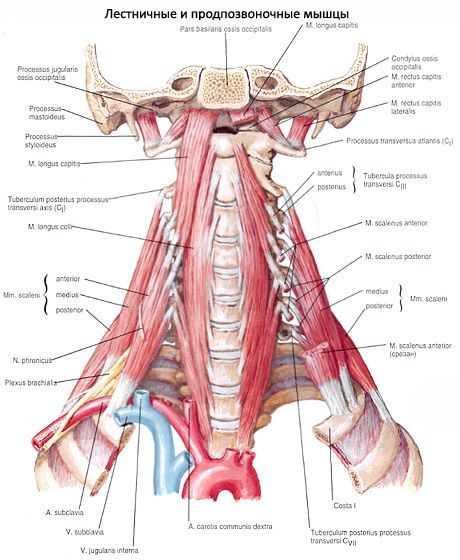
The medial (prevertebral) group of muscles is located on the anterior surface of the spinal column on the sides of the midline and is represented by the long muscles of the neck and head, the anterior and lateral rectus muscles of the head.
The longus colli muscle (m.longus colli) is adjacent to the anterolateral surface of the spine along the length from the 3rd thoracic to the 1st cervical vertebrae. This muscle has three parts: vertical, inferior oblique, and superior oblique. The vertical part originates on the anterior surface of the bodies of the upper three thoracic and three lower cervical vertebrae, passes vertically upward, and is attached to the bodies of the 2nd-4th cervical vertebrae. The inferior oblique part originates on the anterior surface of the bodies of the first three thoracic vertebrae and is attached to the anterior tubercles of the 6th-5th cervical vertebrae. The superior oblique part originates on the anterior tubercles of the transverse processes of the 3rd-5th cervical vertebrae, rises upward, and is attached to the anterior tubercle of the 1st cervical vertebra.
Function: flexes the cervical spine. With unilateral contraction, it tilts the neck to its side. With contraction of the superior oblique part, the head turns to the same side, with contraction of the inferior oblique part - to the opposite side.
Innervation: muscular branches of the cervical plexus (CII-CVI).
Blood supply: vertebral, ascending cervical and deep cervical arteries.
The long muscle of the head (m.longus capitis) begins with four tendon bundles on the anterior tubercles of the transverse processes of the VI-III cervical vertebrae, passes upward and medially; attaches to the lower surface of the basilar part of the occipital bone.
Function: tilts the head and cervical spine forward.
Innervation: muscular branches of the cervical plexus (CI-CV).
Blood supply: vertebral and deep cervical arteries.
The anterior rectus capitis muscle (m.rectus capitis anterior) is located deeper than the longus capitis muscle. It begins on the anterior arch of the atlas and attaches to the basilar part of the occipital bone, posterior to the attachment site of the longus capitis muscle.
Function: tilts the head forward.
Innervation: muscular branches of the cervical plexus (CI-CII).
Blood supply: vertebral and ascending pharyngeal arteries.
The lateral rectus capitis muscle (m.rectus capitis lateralis) is located outside the anterior rectus capitis muscle, begins on the transverse process of the atlas, passes upward and is attached to the lateral part of the occipital bone.
Function: tilts the head to one side, acts exclusively on the atlanto-occipital joint.
Innervation: muscular branches of the cervical plexus (CI).
Blood supply: occipital and vertebral arteries.
 [ 1 ]
[ 1 ]
Where does it hurt?
What do need to examine?