Medical expert of the article

New publications
The conduction system of the heart
Last reviewed: 06.07.2025

All iLive content is medically reviewed or fact checked to ensure as much factual accuracy as possible.
We have strict sourcing guidelines and only link to reputable media sites, academic research institutions and, whenever possible, medically peer reviewed studies. Note that the numbers in parentheses ([1], [2], etc.) are clickable links to these studies.
If you feel that any of our content is inaccurate, out-of-date, or otherwise questionable, please select it and press Ctrl + Enter.
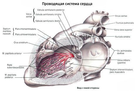
Regulation and coordination of the contractile function of the heart is carried out by its conduction system. The conduction system of the heart is formed by atypical cardiomyocytes (cardiac conduction cardiomyocytes). These cardiomyocytes are richly innervated, have small sizes (length - about 25 μm, thickness - 10 μm) compared to myocardial cardiomyocytes.
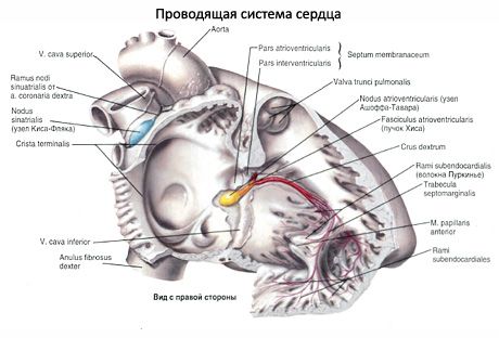
The cells of the conduction system do not have T-tubes, they are connected to each other not only by their ends, but also by their lateral surfaces. These cells contain a significant amount of cytoplasm and few myofibrils. The cells of the conduction system have the ability to conduct irritation from the nerves of the heart to the myocardium of the atria and ventricles. The centers of the conduction system of the heart are two nodes:
- the sinoatrial node (Keith-Fleck node; nodus sinuatrialis) is located in the wall of the right atrium, between the opening of the superior vena cava and the right auricle; it gives off branches to the myocardium of the atria;
- The atrioventricular node (Aschoff-Tawara node; nodus atrioventricularis) lies in the thickness of the lower part of the interatrial septum. Inferiorly, this node passes into the atrioventricular bundle (bundle of His; fasciculus atrioventricularis), which connects the myocardium of the atria with the myocardium of the ventricles. In the muscular part of the interventricular septum, this bundle divides into the right and left crus (crus dextrum et crus sinistrum). The terminal branches of the fibers of the cardiac conduction system (Purkinje fibers), into which these crus are divided, end in the myocardium on the cardiomyocytes of the ventricles.
 [
[