Medical expert of the article

New publications
Blood and lymphatic vessels of the heart
Last reviewed: 04.07.2025

All iLive content is medically reviewed or fact checked to ensure as much factual accuracy as possible.
We have strict sourcing guidelines and only link to reputable media sites, academic research institutions and, whenever possible, medically peer reviewed studies. Note that the numbers in parentheses ([1], [2], etc.) are clickable links to these studies.
If you feel that any of our content is inaccurate, out-of-date, or otherwise questionable, please select it and press Ctrl + Enter.
The arteries of the heart branch off from the aortic bulb, the initial widened section of the ascending aorta. These arteries surround the heart like a crown, which is why they are called the coronary arteries. The right coronary artery begins at the level of the right aortic sinus, and the left one at the level of the left aortic sinus. Both arteries branch off from the aorta below the free (upper) edges of the semilunar valves, so during contraction (systole) of the ventricles, the valves cover the openings of the arteries and almost do not allow blood to pass to the heart. During relaxation (diastole) of the ventricles, the sinuses fill with blood, blocking its path from the aorta back to the left ventricle. At the same time, blood access to the vessels of the heart opens.
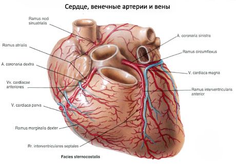
The right coronary artery (a.coronaria dextra) goes to the right under the right atrium appendage, lies in the coronary groove, and bends around the right (pulmonary) surface of the heart. Then the artery follows the posterior surface of the heart to the left, where its end anastomoses with the circumflex branch of the right coronary artery. The largest branch of the right coronary artery is the posterior interventricular branch (r.interventricularis posterior), which goes along the groove of the heart of the same name towards its apex. The branches of the right coronary artery supply blood to the walls of the right ventricle and atrium, the posterior part of the interventricular septum, the papillary muscles of the right ventricle, the posterior papillary muscle of the left ventricle, the sinoatrial and atrioventricular nodes of the cardiac conduction system.
The left coronary artery (a.coronaria sinistra) is somewhat thicker than the right one, located between the beginning of the pulmonary trunk and the appendage of the left atrium. It divides into two branches: the anterior interventricular branch (r.interventricularis anterior) and the circumflex branch (r.circumflexus). The latter, which is a continuation of the main trunk of the coronary artery, bends around the heart on the left, located in its coronary groove. On the posterior side of the organ, this branch anastomoses with the right coronary artery. The anterior interventricular branch follows the groove of the heart of the same name towards its apex. In the area of the cardiac notch, this branch sometimes passes to the diaphragmatic surface of the heart, where it anastomoses with the terminal section of the posterior interventricular branch of the right coronary artery. Branches of the left coronary artery supply the walls of the left ventricle, including the papillary muscles, most of the interventricular septum, the anterior wall of the right ventricle, and the walls of the left atrium.
The branches of the right and left coronary arteries, joining together, form two arterial rings around the heart: a transverse one in the coronary groove and a longitudinal one, the vessels of which are located in the anterior and posterior interventricular grooves.
The branches of the coronary arteries provide blood supply to all layers of the heart walls. In the myocardium, where the level of oxidative processes is highest, the microvessels anastomosing with each other repeat the course of the muscle bundles.
There are different types of distribution of the branches of the coronary arteries, which are called types of blood supply to the heart. The main ones are as follows:
- right coronary type - most parts of the heart are supplied with blood by branches of the right coronary artery;
- left coronary type - most of the heart receives blood from the branches of the left coronary artery;
- average, or uniform, type - both coronary arteries are evenly distributed in the walls of the heart.
Transitional types of blood supply to the heart are also distinguished - middle-right and middle-left. It is generally accepted that among all types of blood supply to the heart, the middle-right type is predominant.
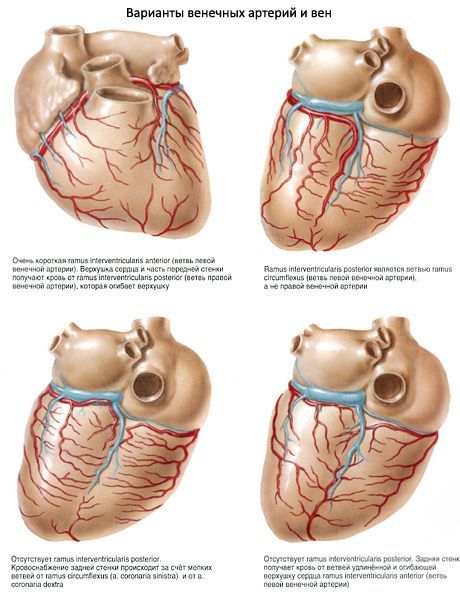
Variations and anomalies of the position and branching of the coronary arteries are possible. They manifest themselves in a change in the place of origin and number of coronary arteries. Thus, the arteries can branch off from the aorta directly above the semilunar valves or significantly higher - from the left subclavian artery, and not from the aorta. The coronary artery can be single, i.e. unpaired; there can be 3-4 coronary arteries, and not two: two arteries branch off to the right and left of the aorta, or two from the aorta and two from the left subclavian artery.
Along with the coronary arteries, non-permanent (additional) arteries go to the heart (especially to the pericardium). These may be the mediastinal-pericardial branches (upper, middle and lower) of the internal thoracic artery, branches of the pericardial diaphragmatic artery, as well as branches extending from the concave surface of the aortic arch, etc.
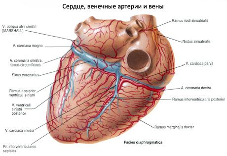
The veins of the heart are more numerous than the arteries. Most of the large veins of the heart gather into one common wide venous vessel - the coronary sinus (sinus coronarius) - a remnant of the embryonic left common cardiac vein. The sinus is located in the coronary groove on the posterior surface of the heart and opens into the right atrium below and in front of the opening of the inferior vena cava (between its valve and the interatrial septum). The tributaries of the coronary sinus are 5 veins:
- the great cardiac vein (v.cardiaca magna), which begins in the region of the apex of the heart on its anterior surface. This vein lies in the anterior interventricular groove next to the anterior interventricular branch of the left coronary artery. Then, at the level of the coronary groove, the vein turns to the left, passes under the circumflex branch of the left coronary artery, lies in the coronary groove on the posterior surface of the heart, where it continues into the coronary sinus. The great cardiac vein collects blood from the veins of the anterior surface of both ventricles and the interventricular septum. The veins of the posterior surface of the left atrium and left ventricle also flow into the great cardiac vein;
- the middle cardiac vein (v.cardiaca media) is formed in the area of the posterior surface of the apex of the heart, rises up along the posterior interventricular groove (adjacent to the posterior interventricular branch of the right coronary artery) and flows into the coronary sinus;
- the small cardiac vein (v.cardiaca parva) begins on the right (pulmonary) surface of the right ventricle, rises upward, lies in the coronary groove on the diaphragmatic surface of the heart and flows into the coronary sinus. This vein collects blood mainly from the right half of the heart;
- the posterior vein of the left ventricle (v.posterior ventriculi sinistri) is formed from several veins on the posterior surface of the left ventricle, closer to the apex of the heart and flows into the coronary sinus or into the great vein of the heart;
- The oblique vein of the left atrium (v.obhqua atrii sinistri) runs from top to bottom along the posterior surface of the left atrium and flows into the coronary sinus.
In addition to the veins that flow into the coronary sinus, the heart has veins that open directly into the right atrium. These are the anterior cardiac veins (vv.cardiacae anteriores), which collect blood from the anterior wall of the right ventricle. They run upward to the base of the heart and open into the right atrium. The smallest cardiac veins (thebesian veins; vv.cardiacae minimae), 20-30 in total, begin in the thickness of the heart walls and flow directly into the right atrium and partly into the ventricles and left atrium through the openings of the smallest veins.
The lymphatic bed of the heart walls consists of lymphatic capillaries located in the form of networks in the endocardium, myocardium and epicardium. Lymph from the endocardium and myocardium flows into the superficial network of lymphatic capillaries located in the epicardium and into the plexus of lymphatic vessels. Connecting with each other, the lymphatic vessels enlarge and form two main vessels of the heart, through which the lymph flows to the regional lymph nodes. The left lymphatic vessel of the heart is formed from the confluence of the lymphatic vessels of the anterior surfaces of the right and left ventricles, the left pulmonary and posterior surfaces of the left ventricle. It follows from the left ventricle to the right, passes behind the pulmonary trunk and flows into one of the lower tracheobronchial lymph nodes. The right lymphatic vessel of the heart is formed from the lymphatic vessels of the anterior and posterior surfaces of the right ventricle, is directed from right to left along the anterior semicircle of the pulmonary trunk and flows into one of the anterior mediastinal lymph nodes located at the arterial ligament. Small lymphatic vessels, through which lymph flows from the walls of the atria, flow into nearby anterior mediastinal lymph nodes.
 [
[