Medical expert of the article

New publications
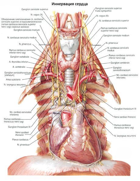
The nerves of the heart
Last reviewed: 06.07.2025

All iLive content is medically reviewed or fact checked to ensure as much factual accuracy as possible.
We have strict sourcing guidelines and only link to reputable media sites, academic research institutions and, whenever possible, medically peer reviewed studies. Note that the numbers in parentheses ([1], [2], etc.) are clickable links to these studies.
If you feel that any of our content is inaccurate, out-of-date, or otherwise questionable, please select it and press Ctrl + Enter.
The heart receives sensory, sympathetic and parasympathetic innervation. Sympathetic fibers, which go as part of the cardiac nerves from the right and left sympathetic trunks, carry impulses that accelerate the heart rate and expand the lumen of the coronary arteries. Parasympathetic fibers (a component of the cardiac branches of the vagus nerves) conduct impulses that slow the heart rate and narrow the lumen of the coronary arteries. Sensory fibers from the receptors of the walls of the heart and its vessels go as part of the cardiac nerves and cardiac branches to the corresponding centers of the spinal cord and brain.
The diagram of the innervation of the heart (according to V.P. Vorobyov) can be presented as follows. The cardiac nerves and branches that go to the heart form extraorgan cardiac plexuses (superficial and deep), located near the aortic arch and pulmonary trunk. The intraorgan cardiac plexus is located in the walls of the heart and is distributed in all their layers.
The cardiac (sympathetic) nerves (upper, middle and lower cervical, as well as thoracic) originate from the cervical and upper thoracic (II and V) nodes of the right and left sympathetic trunks (see "Autonomic nervous system"). The cardiac branches originate from the right and left vagus nerves (see "Vagus nerve").
The superficial extraorgan cardiac plexus lies on the anterior surface of the pulmonary trunk and on the concave semicircle of the aortic arch. The deep extraorgan cardiac plexus is located behind the aortic arch (in front of the tracheal bifurcation). The superficial extraorgan cardiac plexus receives the upper left cervical cardiac nerve (from the left upper cervical sympathetic ganglion) and the upper left cardiac branch (from the left vagus nerve). All other named cardiac nerves and cardiac branches enter the deep extraorgan cardiac plexus.
The branches of the extraorgan cardiac plexuses pass into a single intraorgan cardiac plexus. Depending on which layer of the heart wall it is located in, this cardiac plexus is conventionally divided into closely interconnected subepicardial, intramuscular and subendocardial plexuses. The intraorgan cardiac plexus contains nerve cells and their clusters that belong to the parasympathetic part of the autonomic nervous system and form small-sized cardiac nerve nodules (ganglia cardiaca). There are especially many nerve cells in the subepicardial cardiac plexus. According to V.P. Vorobyov, the nerves that make up the subepicardial cardiac plexus have a regular arrangement (in the form of nodal fields) and innervate certain areas of the heart. Accordingly, six subepicardial cardiac plexuses are distinguished - three on the anterior side of the heart, three on the posterior:
- right front;
- left anterior. They are located under the epicardium of the anterior and lateral walls of the right and left ventricles on both sides of the arterial cone;
- the anterior atrial plexus is localized in the anterior wall of the atria;
- the right posterior plexus descends from the posterior wall of the right atrium into the posterior wall of the right ventricle (fibers go from it to the sinoatrial node of the cardiac conduction system);
- the left posterior plexus from the lateral wall of the left atrium continues downward into the posterior wall of the left ventricle;
- The posterior plexus of the left atrium (plexus of the Hallerian sinus) is located in the upper part of the posterior wall of the left atrium (between the mouths of the pulmonary veins).
 [
[