Medical expert of the article

New publications
Sternoclavicular-papillary muscle.
Last reviewed: 04.07.2025

All iLive content is medically reviewed or fact checked to ensure as much factual accuracy as possible.
We have strict sourcing guidelines and only link to reputable media sites, academic research institutions and, whenever possible, medically peer reviewed studies. Note that the numbers in parentheses ([1], [2], etc.) are clickable links to these studies.
If you feel that any of our content is inaccurate, out-of-date, or otherwise questionable, please select it and press Ctrl + Enter.
The sternocleidomastoid muscle (m. sternocleidomastoideus) is located under the platysma muscle of the neck; when the head is turned to the side, its contour is indicated by a distinct ridge on the anterolateral surface of the neck. This muscle begins with two parts (medial and lateral) on the anterior surface of the manubrium of the sternum and the sternal end of the clavicle. Rising upward and backward, the muscle is attached to the mastoid process of the temporal bone and the lateral segment of the superior nuchal line of the occipital bone. Above the clavicle, between the medial and lateral parts of the muscle, there is a small supraclavicular fossa (fossa supraclavicularis minor).
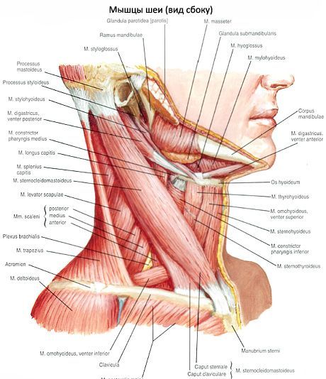
Function of the sternocleidomastoid muscle: with a unilateral contraction, it tilts the head to one side, while simultaneously turning the face to the opposite side. With a bilateral contraction of the muscle, the head is thrown back, since the muscle is attached behind the transverse axis of the atlanto-occipital joint. With a fixed head, the muscle pulls the chest upward, facilitating inhalation, as an auxiliary respiratory muscle.
Innervation of the sternocleidomastoid muscle: accessory nerve (XI).
Blood supply of the sternocleidomastoid muscle: sternocleidomastoid branches of the occipital and superior thyroid arteries.
 [
[ Where does it hurt?
What do need to examine?