Medical expert of the article
New publications
Bowen's disease
Last reviewed: 04.07.2025

All iLive content is medically reviewed or fact checked to ensure as much factual accuracy as possible.
We have strict sourcing guidelines and only link to reputable media sites, academic research institutions and, whenever possible, medically peer reviewed studies. Note that the numbers in parentheses ([1], [2], etc.) are clickable links to these studies.
If you feel that any of our content is inaccurate, out-of-date, or otherwise questionable, please select it and press Ctrl + Enter.

Bowen's disease (syn.: squamous cell carcinoma in situ, intraepidermal cancer) is a rare typical variant of non-invasive cancer, which often appears on sun-exposed skin areas. This type of cancer usually develops in older people. The exact cause of development is unknown, although some risk factors have been identified. The lesions are usually painless. Treatment is usually surgical. The prognosis of the disease is favorable.
Epidemiology
The prevalence of the disease varies by region and ranges from 14.9 cases per 100,000 to 142 cases per 100,000.
There is no difference between the incidence of the disease in men and women. It most often develops in adulthood, with a high frequency in patients over 60 years of age.
Causes Bowen's disease
The exact cause of the disease is unknown.
Risk factors
Like other forms of skin cancer, Bowen's disease develops as a result of chronic sun exposure and aging. Oncogenic papillomavirus (HPV 16, 2, 34, 35) and chronic arsenic intoxication are also considered to be causes of the disease.
People with fair skin who spend a lot of time in direct sunlight, people taking cytostatic drugs, organ transplant patients, and HIV-infected people are at high risk of developing this disease.
Pathogenesis
Acanthosis with elongation and thickening of epidermal outgrowths, hyperkeratosis, focal parakeratosis are revealed. The basal layer is without any significant changes. Spinous cells are located randomly, many of them with sharply expressed atypia of large hyperchromic nuclei. Large multinuclear cells with intensely stained nuclei are often found, mitotic figures are encountered. Foci of dyskeratosis are formed from large rounded cells with homogeneous eosinophilic cytoplasm and pycnotic nucleus. Sometimes foci of incomplete keratinization can be found in the form of concentric layers of keratinized cells, resembling "horny pearls". Some cells are strongly vacuolated, similar to Paget's cells, but the latter do not have intercellular bridges. When Bowen's disease progresses to invasive cancer, acanthotic cords penetrate deeply into the dermis with disruption of the basement membrane and pronounced polymorphism of the cells in these cords.
Symptoms Bowen's disease
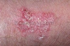
Characterized by the presence of a usually solitary, sharply limited lesion, round or oval outlines, less often irregular in shape, with slow peripheral growth with the formation of a slightly raised edge, flaky or covered with crusts. The surface is uneven, granular, and may be slightly warty. Superficial erosion, ulceration with the formation of partially scarring and at the same time increasing ulcers on the surface are observed. Most often, lesions are located on the head, hands, genitals, but can be on any part of the skin and on the mucous membranes. With a long course, transformation into typical squamous cell carcinoma may occur.
What do need to examine?
How to examine?
Differential diagnosis
Bowen's disease should be differentiated from seborrheic keratosis, in which pigmentation and intraepidermal cysts are often expressed, the cells are darker and smaller, and their atypia is less pronounced.
Treatment Bowen's disease
Treatment depends on each individual case and depends on many factors, such as:
- location, size and thickness of the pathological lesion;
- the presence or absence of certain symptoms;
- age and general health.
Photodynamic therapy (PDT), cryotherapy, local chemotherapy with 5-fluorouracil are used in the treatment. Recent studies (2013) have shown good efficiency of using 5% Imiquimod cream in local therapy. As a rule, the cream is applied once or twice a day for at least two weeks.
Cryotherapy is most effective for single and small lesions.
Some dermatologists prefer to perform surgical intervention by excising the pathological lesion.


 [
[