Medical expert of the article

New publications
Seborrheic keratosis: causes, symptoms, diagnosis, treatment
Last reviewed: 04.07.2025

All iLive content is medically reviewed or fact checked to ensure as much factual accuracy as possible.
We have strict sourcing guidelines and only link to reputable media sites, academic research institutions and, whenever possible, medically peer reviewed studies. Note that the numbers in parentheses ([1], [2], etc.) are clickable links to these studies.
If you feel that any of our content is inaccurate, out-of-date, or otherwise questionable, please select it and press Ctrl + Enter.
Seborrheic keratosis (syn.: seborrheic wart, keratoma, senile wart, basal cell papilloma, seborrheic nevus of Unna, seborrheic keratopapilloma) is a benign tumor. A fairly common disease that occurs mainly in the second half of life, less often - at a younger age.
Here is some information about seborrheic keratoses:
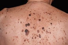
- Appearance: Localized on the face, trunk. It is a sharply defined hyperpigmented spot with a smooth or slightly scaly surface up to several centimeters in diameter, or a plaque-like or nodular formation with a warty surface and varying degrees of pigmentation, covered with dry horny masses. It can be single, more often multiple. They can be of different colors, including brown, black, white and even pink. The surface of seborrheic keratoses can be rough and often has a texture similar to a wax wallet or walnut.
- Distribution: Seborrheic keratoses most often appear on areas of the skin that are exposed to the sun, such as the face, chest, back, neck, and hands. However, they can appear in other places as well.
- Symptoms: Seborrheic keratoses usually do not cause pain or discomfort. They may become noticeable to the touch, but are not usually associated with itching or soreness.
- Treatment: Most cases do not require treatment unless they cause cosmetic or physical problems. If the growth is bothersome, it can be removed using surgical methods such as electrocoagulation, cryotherapy (freezing), laser removal or excision.
- Prevention: To prevent new tumors from developing and reduce the risk of skin cancer, it is important to use sunscreen, wear protective clothing, and avoid prolonged exposure to the sun.
- Consult a Doctor: If you notice changes in your skin or have new growths, it is best to consult a dermatologist. The doctor will be able to diagnose and give recommendations for skin care.
Causes seborrheic keratoma
The causes of seborrheic keratoses are not fully understood, but they are thought to be related to age and genetics. Here are some factors that may play a role in the development of seborrheic keratosis:
- Age: Seborrheic keratoses most often occur in people over 40-50 years of age. The likelihood of their occurrence increases with age.
- Genetics: Genetics may play a role. If family members have had these tumors, you may also have an increased risk of developing them.
- Sun exposure: Prolonged and repeated exposure to ultraviolet (UV) radiation from the sun can contribute to the development of these growths. Therefore, they are more common on sun-exposed skin.
- Hormonal changes: Some studies have shown that hormonal changes, such as pregnancy or hormone replacement therapy, may influence the development of seborrheic keratoses.
- Skin conditions: People with certain skin conditions, such as xeroderma pigmentosum, may develop seborrheic keratoses in greater numbers.
It is important to note that seborrheic keratoses are benign growths and rarely develop into cancer. However, if you have new or changing skin growths, it is important to consult a doctor for diagnosis and monitoring.
Pathogenesis
Pathomorphology. Seborrheic keratosis mostly has an exophytic papillomatous growth type, less often with spreading into the dermis in the form of massive layers of epithelial cells of various configurations. Histologically, "irritated" (hyperkeratotic), adenoid or reticular, flat (acantotic) types of seborrheic keratosis are distinguished. Often, the same lesion can combine signs of all types.
The hyperkeratotic type is characterized by acanthosis, hyperkeratosis, and papillomatosis. The stratum corneum invaginates into the epidermis in places, resulting in the formation of cystic cavities filled with horny masses (pseudo-horny cysts). The acanthotic cords consist mainly of spinous cells, but in places there are clusters of basaloid cells.
The flat (acanthotic) type is characterized by a sharp thickening of the epidermis with relatively moderate hyperkeratosis and papillomatosis. There are a large number of pseudo-horny cysts with a predominance of basaloid cells along the periphery.
In the adenoid type, there is a proliferation of numerous narrow branching strands consisting of 1-2 rows of basaloid cells in the upper parts of the dermis. Horny cysts are sometimes of significant size, in connection with which we can talk about the alenoid-cystic variant.
In the "irritated" type of seborrheic keratosis, a significant inflammatory infiltrate is detected in the dermis with exocytosis of the cellular elements of the infiltrate into the structures of the neoplasm, which is accompanied by squamous epithelial differentiation and the formation of numerous rounded keratinization foci, designated in the English-language literature as eddies. The histological picture in these cases is similar to that of pseudoepitheliomatous hyperplasia or follicular keratoma.
MR Qtaffl and LM Edelstem (1976) identified the so-called clonal type of seborrheic keratosis, which has intraepidermal proliferations of basaloid cells. The clonal type of seborrheic keratosis can arise as a result of exogenous influences and is characterized by the transformation of basaloid cells into spinous ones. Clearly delimited complexes of small monomorphic basaloid cells can form intraepithelially, like the so-called Burst-Jadasson epithelioma. Finally, some authors identify a superficial type of multiple papillomatous keratomas with signs of seborrheic teratoma - stuccokeratose, in which hyperkeratosis in the form of "church spires" is noted. Seborrheic keratoma cells are small polygonal, with dark oval nuclei, and resemble basal cells of the epidermis, which is reflected in the name of one of the synonyms. Among these cells are horny cysts, near which one can see the transition of basaloid cells into spiny cells with keratinization phenomena. Horny cysts can also be found in deeper parts of the acanthotic cords.
Seborrheic keratoma cells may contain varying amounts of pigment, which ultimately determines the color of the tumor element itself. Lymphohistiocytic or plasma cell infiltrates are often found in the stroma of seborrheic keratoma.
Histogenesis. Electron microscopy revealed that basaloid cells can originate from both spinous and basal cells and are distinguished by a high density of cytoplasm. They have fewer tonofilaments, but their orientation is the same as that of normal epidermis cells, and there is a sufficient number of desmosomes. A. Ackerman et al. (1993) provide information on the histogenetic commonality of seborrheic and follicular keratosis, linking their genesis with the cells of the epithelial lining of the infundibulum of the hair follicle. Studies devoted to the study of intraepidermal macrophages, which may be regulators of the keratinization processes of epithelial cells, have shown that there are significantly more of them in seborrheic keratosis than in normal skin.
Multiple seborrheic keratomas are observed in Leser-Trelat syndrome, and their number rapidly increases in malignant tumors of internal organs, especially the stomach.
Differentiation with early stages of squamous cell carcinoma and precancerous actinic keratosis is very difficult. The most important feature in these cases is horny or pseudo-horny cysts, the absence of cellular atypia around them and the presence of basaloid cells on the periphery. In the eccrine poroma, which in its histological structure can be very similar to seborrheic keratoma of solid structure, there are duct structures, the cells contain glycogen, horny cysts and pigment are absent.
Symptoms seborrheic keratoma
Seborrheic keratoses usually have characteristic signs and symptoms that can vary depending on their size, color, and location on the skin. Here are the main symptoms:
- Appearance: Seborrheic keratoses appear as flat or slightly raised wart-like spots or growths on the skin. They can range in size from a few millimeters to several centimeters. The surface of the keratomas is often rough and has a texture reminiscent of a wax wallet or walnut.
- Color: May come in a variety of colors, including brown, black, white, yellow, and pink. Color may vary depending on individual characteristics.
- Distribution: These growths can appear on different areas of the skin, but are most common on sun-exposed areas such as the face, chest, back, neck, and arms. However, they can appear in other areas as well.
- No symptoms: These growths usually do not cause pain or discomfort. They may be noticeable to the touch, but are not usually associated with itching, redness, or tenderness.
- Number: A person may have multiple tumors, and the number may increase with age.
These growths are usually benign and rarely require treatment unless they cause cosmetic or physical concerns. However, it is important to monitor skin growths for changes and consult a doctor if new or changing growths appear.
Diagnostics seborrheic keratoma
Diagnosis of seborrheic keratosis is usually made visually by examining the skin by a doctor, most often a dermatologist. The examination can identify the characteristic signs of this skin problem. In some cases, the following diagnostic methods may be required to rule out other diseases or more serious skin changes:
- Dermoscopy: Dermoscopy is a technique that uses a special magnifying device called a dermoscope to examine the skin's structure in more detail. This technique can help your doctor identify the characteristic features of a tumor and differentiate it from other skin growths.
- Biopsy: In rare cases, your doctor may decide to perform a biopsy, in which a small sample of tissue from the seborrheic keratosis is removed and sent to a lab for testing. This helps rule out the possibility that the growth is cancerous.
- Clinical evaluation: The doctor may also ask questions about the patient's symptoms and medical history to get a better understanding of the skin condition.
What do need to examine?
How to examine?
Who to contact?
Treatment seborrheic keratoma
Seborrheic keratoses generally do not require treatment because they are benign and do not cause physical discomfort or health risks. However, sometimes patients may want to remove a seborrheic keratoma for cosmetic reasons or because of discomfort caused by their location. Here are some removal methods:
- Cryotherapy: This method uses liquid nitrogen to freeze and remove the tumor. The growth usually falls off on its own within a few days after the procedure.
- Electrocautery: The doctor uses an electric current to burn away the growth. This method can be effective but leaves small scars.
- Laser removal: Laser removal can be painless and leaves less scarring. The laser is used to vaporize the top layer of the keratosis.
- Surgical removal: In some cases, your doctor may decide to perform surgical removal. This may require stitches and leave scars.
- Chemical removal: The doctor may use chemical agents, such as acids, to remove seborrheic keratosis. This method may also require multiple sessions.
It is important to consult a dermatologist or other specialist to determine the most appropriate method for removing the tumor, taking into account its size, number, and location on the skin. It is not recommended to try to remove the growth yourself without consulting a doctor, as this may lead to infection or complications.
Prevention
Seborrheic keratoses are usually caused by natural skin aging and genetic predisposition, and preventing them from occurring is almost impossible. However, there are some steps you can take to reduce the risk of them occurring or to slow down the process:
- UV protection: Avoid prolonged exposure to sunlight and use sunscreens with a high SPF, wear skin-protective clothing and wear wide-brimmed hats.
- Skin Care: Take regular care of your skin with moisturizers and creams to keep it healthy and supple.
- Avoid trauma and friction: Seborrheic keratoses can sometimes occur in areas that are subject to constant friction or trauma. Try to avoid such situations.
- Maintaining a Healthy Lifestyle: Eating a healthy diet and avoiding unhealthy habits can contribute to overall skin health.
- Regular check-ups with a dermatologist: It is important to have regular check-ups with a specialist to detect and monitor any changes in skin lesions. This will help to identify and treat any skin problems in a timely manner.
Forecast
The prognosis for seborrheic keratoses is usually very good. These skin growths are benign and rarely pose any health risks. They are not associated with the development of cancer or other serious diseases.
Seborrheic keratoses can appear on the skin over time and may increase in size and number with age. They may be removed if they cause cosmetic discomfort, but this is usually not medically necessary.
It is important to monitor skin lesions for changes and see a doctor if you notice any unusual symptoms, such as a sudden increase in size, change in color, bleeding, itching, or pain.
Some classic books and authors in the field of oncology that may be helpful
- "Cancer: Principles & Practice of Oncology" (Book on the principles and practice of oncology) - Authors: Vincent T. DeVita Jr., Theodore S. Lawrence, Steven A. Rosenberg, et al.
- "The Emperor of All Maladies: A Biography of Cancer" - By Siddhartha Mukherjee
- "Oxford Textbook of Oncology" - Authors: David J. Kerr, Daniel G. Haller, Cornelis JH van de Velde and others.
- "Principles and Practice of Gynecologic Oncology" - Authors: Dennis S. Chi, Andrew Berchuck, Robert L. Coleman, et al.
- "The Biology of Cancer" - Author: Robert A. Weinberg
- "Clinical Oncology" - Authors: Martin D. Abeloff, James O. Armitage, John E. Niederhuber, et al.
- "Oncology: An Evidence-Based Approach" - Authors: Alfred E. Chang, Patricia A. Ganz, Daniel F. Hayes, et al.
References
- Chissov, V. I. Oncology: National Guide. Brief edition / edited by V. I. Chissov, M. I. Davydov - Moscow: GEOTAR-Media, 2017.
- Butov, Yu. S. Dermatovenereology. National leadership. Brief edition / ed. Yu. S. Butova, Yu. K. Skripkina, O. L. Ivanova. - Moscow: GEOTAR-Media, 2020.