Medical expert of the article

New publications
Biomicroscopy
Last reviewed: 07.07.2025

All iLive content is medically reviewed or fact checked to ensure as much factual accuracy as possible.
We have strict sourcing guidelines and only link to reputable media sites, academic research institutions and, whenever possible, medically peer reviewed studies. Note that the numbers in parentheses ([1], [2], etc.) are clickable links to these studies.
If you feel that any of our content is inaccurate, out-of-date, or otherwise questionable, please select it and press Ctrl + Enter.
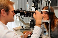
Biomicroscopy is intravital microscopy of eye tissues, a method that allows examining the anterior and posterior sections of the eyeball under different lighting and image sizes. The study is conducted using a special device - a slit lamp, which is a combination of an illumination system and a binocular microscope. Using a slit lamp, you can see the details of the tissue structure in a living eye. The illumination system includes a slit-shaped diaphragm, the width of which can be adjusted, and filters of different colors. A beam of light passing through the slit forms a light section of the optical structures of the eyeball, which is examined through a slit-lamp microscope. By moving the light slit, the doctor examines all the structures of the anterior section of the eye.
The patient's head is placed on a special slit lamp stand with the chin and forehead supported. The illuminator and microscope are moved to the patient's eye level. The light slit is alternately focused on the tissue of the eyeball that is to be examined. The light beam directed at the translucent tissues is narrowed and the light intensity is increased to obtain a thin light section. In the optical section of the cornea, it is possible to see foci of opacities, newly formed vessels, infiltrates, assess their depth, and identify various tiny deposits on its posterior surface. When examining the marginal looped vascular network and conjunctival vessels, it is possible to observe the blood flow in them and the movement of blood cells.
Biomicroscopy allows one to clearly examine various zones of the lens (anterior and posterior poles, cortex, nucleus), and if its transparency is impaired, to determine the localization of pathological changes. The anterior layers of the vitreous body are visible behind the lens.
There are four different methods of biomicroscopy depending on the type of lighting:
- in direct focused light, when the light beam of a slit lamp is focused on the area of the eyeball being examined. This allows one to assess the degree of transparency of optical media and identify areas of opacities;
- in reflected light. This allows one to examine the cornea in rays reflected from the iris when searching for foreign bodies or identifying areas of swelling;
- in indirect focused light, when the light beam is focused near the area being examined, which allows for better visibility of changes due to the contrast between highly and poorly illuminated areas;
- with indirect diaphragmatic transillumination, when reflective (mirror) zones are formed at the interface between optical media with different refractive indices of light, which allows one to examine areas of tissue near the point where the reflected beam of light emerges (examination of the anterior chamber angle).
With the specified types of lighting, two techniques can also be used:
- conduct a study in a sliding beam (when the handle of the slit lamp moves the light strip along the surface left and right), which allows you to detect unevenness of the relief (corneal defects, newly formed vessels, infiltrates) and determine the depth of these changes;
- perform research in a mirror field, which also helps to study the surface relief and at the same time identify irregularities and roughness.
The use of additional aspherical lenses (such as the Gruby lens) during biomicroscopy makes it possible to perform ophthalmoscopy of the fundus (against the background of drug-induced mydriasis), revealing subtle changes in the vitreous body, retina and choroid.
Modern design and adaptations of slit lamps also allow for additional determination of the thickness of the cornea and its external parameters, assessment of its reflectivity and sphericity, and measurement of the depth of the anterior chamber of the eyeball.
An important achievement of recent years is ultrasound biomicroscopy, which allows one to examine the ciliary body, the posterior surface and section of the iris, and the lateral sections of the lens, which are hidden behind the opaque iris during conventional light biomicroscopy.
 [
[